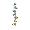
 Movie
Movie Controller


+ Open data

- Basic information
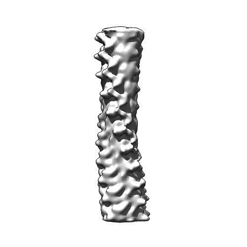
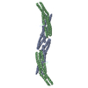
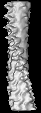
| Entry | Database: EMDB / ID: EMD-2946 | |||||||||
|---|---|---|---|---|---|---|---|---|---|---|
| Title | cryo-EM structure of HET-s prion infectious form | |||||||||
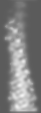
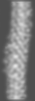
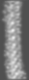
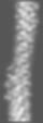
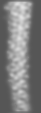
 Map data Map data | Reconstruction of HET-s prion amyloid assembled at pH3 | |||||||||
 Sample Sample |
| |||||||||
 Keywords Keywords | amyloid / prion / cross-beta structure | |||||||||
| Function / homology | Het-s prion-forming domain / Het-s prion-forming domain / Prion-inhibition and propagation, HeLo domain / HeLo domain superfamily / Het-s 218-289 / Prion-inhibition and propagation / identical protein binding / cytoplasm / Heterokaryon incompatibility protein s Function and homology information Function and homology information | |||||||||
| Biological species |  Podospora anserina (fungus) Podospora anserina (fungus) | |||||||||
| Method | helical reconstruction / cryo EM / Resolution: 8.5 Å | |||||||||
 Authors Authors | Mizuno N / Baxa U / Steven AC | |||||||||
 Citation Citation | Journal: Proc Natl Acad Sci U S A / Year: 2011 Title: Structural dependence of HET-s amyloid fibril infectivity assessed by cryoelectron microscopy. Authors: Naoko Mizuno / Ulrich Baxa / Alasdair C Steven /  Abstract: HET-s is a prion protein of the fungus Podospora anserina which, in the prion state, is active in a self/nonself recognition process called heterokaryon incompatibility. Its prionogenic properties ...HET-s is a prion protein of the fungus Podospora anserina which, in the prion state, is active in a self/nonself recognition process called heterokaryon incompatibility. Its prionogenic properties reside in the C-terminal "prion domain." The HET-s prion domain polymerizes in vitro into amyloid fibrils whose properties depend on the pH of assembly; above pH 3, infectious singlet fibrils are produced, and below pH 3, noninfectious triplet fibrils. To investigate the correlation between structure and infectivity, we performed cryo-EM analyses. Singlet fibrils have a helical pitch of approximately 410 Å and a left-handed twist. Triplet fibrils have three protofibrils whose lateral dimensions (36 × 25 Å) and axial packing (one subunit per 9.4 Å) match those of singlets but differ in their supercoiling. At 8.5-Å resolution, the cross-section of the singlet fibril reconstruction is largely consistent with that of a β-solenoid model previously determined by solid-state NMR. Reconstructions of the triplet fibrils show three protofibrils coiling around a common axis and packed less tightly at pH 3 than at pH 2, eventually peeling off. Taken together with the earlier observation that fragmentation of triplet fibrils by sonication does not increase infectivity, these observations suggest a novel mechanism for self-propagation, whereby daughter fibrils nucleate on the lateral surface of singlet fibrils. In triplets, this surface is occluded, blocking nucleation and thereby explaining their lack of infectivity. | |||||||||
| History |
|
- Structure visualization
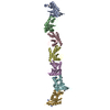
| Movie |
Movie viewer |
|---|---|
| Structure viewer | EM map: SurfViewMolmilJmol/JSmol |
| Supplemental images |
- Downloads & links
-EMDB archive
| Map data | emd_2946.map.gz | 548.5 KB | EMDB map data format | |
|---|---|---|---|---|
| Header (meta data) | emd-2946-v30.xmlemd-2946.xml | 10 KB 10 KB | Display Display | EMDB header |
| Images | 2946-HET-s.tif | 62.9 KB | ||
| Archive directory |  http://ftp.pdbj.org/pub/emdb/structures/EMD-2946ftp://ftp.pdbj.org/pub/emdb/structures/EMD-2946 http://ftp.pdbj.org/pub/emdb/structures/EMD-2946ftp://ftp.pdbj.org/pub/emdb/structures/EMD-2946 | HTTPS FTP |
-Related structure data
| Similar structure data |
|---|


-Links
| EMDB pages | EMDB (EBI/PDBe) / EMDataResource |
|---|---|
| Related items in Molecule of the Month |


-Map
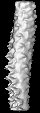
| File | Download / File: emd_2946.map.gz / Format: CCP4 / Size: 708 KB / Type: IMAGE STORED AS FLOATING POINT NUMBER (4 BYTES) | ||||||||||||||||||||||||||||||||||||||||||||||||||||||||||||||||||||
|---|---|---|---|---|---|---|---|---|---|---|---|---|---|---|---|---|---|---|---|---|---|---|---|---|---|---|---|---|---|---|---|---|---|---|---|---|---|---|---|---|---|---|---|---|---|---|---|---|---|---|---|---|---|---|---|---|---|---|---|---|---|---|---|---|---|---|---|---|---|
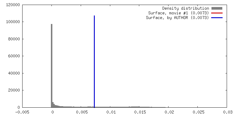
| Annotation | Reconstruction of HET-s prion amyloid assembled at pH3 | ||||||||||||||||||||||||||||||||||||||||||||||||||||||||||||||||||||
| Projections & slices | Image control
Images are generated by Spider. generated in cubic-lattice coordinate | ||||||||||||||||||||||||||||||||||||||||||||||||||||||||||||||||||||
| Voxel size | X=Y=Z: 1.84 Å | ||||||||||||||||||||||||||||||||||||||||||||||||||||||||||||||||||||
| Density |
| ||||||||||||||||||||||||||||||||||||||||||||||||||||||||||||||||||||
| Symmetry | Space group: 1 | ||||||||||||||||||||||||||||||||||||||||||||||||||||||||||||||||||||
| Details | EMDB XML:
CCP4 map header:
| ||||||||||||||||||||||||||||||||||||||||||||||||||||||||||||||||||||
 Z (Sec.)
 Y (Row.)
 X (Col.)


-Supplemental data
- Sample components
-Entire : HET-s prion domain (218-289) assembled at pH3
| Entire | Name: HET-s prion domain (218-289) assembled at pH3 |
|---|---|
| Components |
|
-Supramolecule #1000: HET-s prion domain (218-289) assembled at pH3
| Supramolecule | Name: HET-s prion domain (218-289) assembled at pH3 / type: sample / ID: 1000 Oligomeric state: helical polymer with a rise of 9.4 A per protein unit Number unique components: 1 |
|---|---|
| Molecular weight | Experimental: 8 KDa |
-Macromolecule #1: Heterokaryon incompatibility protein s
| Macromolecule | Name: Heterokaryon incompatibility protein s / type: protein_or_peptide / ID: 1 / Name.synonym: HET-s Details: HET-s proteins are assembled to make a single strand helix, with a rise of 9.4 A per protein. Oligomeric state: Helical filament / Recombinant expression: Yes |
|---|---|
| Source (natural) | Organism: Podospora anserina (fungus) |
| Molecular weight | Experimental: 8 KDa / Theoretical: 8 KDa |
| Recombinant expression | Organism:  |
| Sequence | UniProtKB: Heterokaryon incompatibility protein s / InterPro: Het-s prion-forming domain |
-Experimental details
-Structure determination
| Method | cryo EM |
|---|---|
 Processing Processing | helical reconstruction |
| Aggregation state | filament |
-Sample preparation
| Concentration | 1 mg/mL |
|---|---|
| Buffer | pH: 3 / Details: 20 mM citrate acid |
| Grid | Details: Glow discharged Quantifoil R2/2 was used. |
| Vitrification | Cryogen name: ETHANE / Chamber humidity: 90 % / Instrument: FEI VITROBOT MARK II / Method: Blot for 5 seconds before plunging |
- Electron microscopy
| Microscope | FEI/PHILIPS CM200FEG |
|---|---|
| Temperature | Min: 90 K / Max: 100 K |
| Alignment procedure | Legacy - Astigmatism: Objective lens astigmatism was corrected at 120,000 times magnification |
| Date | Apr 1, 2008 |
| Image recording | Category: FILM / Film or detector model: KODAK SO-163 FILM / Digitization - Scanner: ZEISS SCAI / Digitization - Sampling interval: 7 µm / Number real images: 100 / Average electron dose: 20 e/Å2 / Bits/pixel: 8 |
| Electron beam | Acceleration voltage: 120 kV / Electron source:  FIELD EMISSION GUN FIELD EMISSION GUN |
| Electron optics | Illumination mode: FLOOD BEAM / Imaging mode: BRIGHT FIELD / Cs: 2 mm / Nominal defocus max: 2.5 µm / Nominal defocus min: 1.0 µm / Nominal magnification: 38000 |
| Sample stage | Specimen holder model: GATAN LIQUID NITROGEN |
-Image processing
| Details | The reconstruction was performed using SPIDER in combination with IHRSR. |
|---|---|
| Final reconstruction | Applied symmetry - Helical parameters - Δz: 18.8 Å Applied symmetry - Helical parameters - Δ&Phi: 17.1 ° Applied symmetry - Helical parameters - Axial symmetry: C1 (asymmetric) Algorithm: OTHER / Resolution.type: BY AUTHOR / Resolution: 8.5 Å / Resolution method: OTHER / Software - Name: SPIder, EMAN, IHRSR, bsoft |
| CTF correction | Details: Each micrograph |
