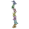
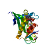
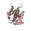
Deposited unit
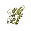
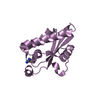
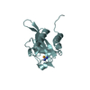
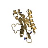
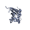
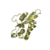
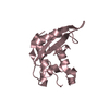
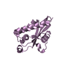
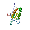
A: PR-1 protein
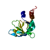
B: PR-1 protein
C: PR-1 protein
D: PR-1 protein
E: PR-1 protein
F: PR-1 protein
G: PR-1 protein
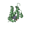
hetero molecules Summary Component details
Theoretical mass Number of molelcules Total (without water) 138,303 16 Polymers 137,196 7 Non-polymers 1,107 9 Water 0 0
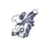
1
A: PR-1 protein
hetero molecules Summary Component details Symmetry operations Calculated values
Theoretical mass Number of molelcules Total (without water) 19,722 2 Polymers 19,599 1 Non-polymers 123 1 Water 0
Type Name Symmetry operation Number identity operation 1_555 x,y,z 1
2
B: PR-1 protein
hetero molecules Summary Component details Symmetry operations Calculated values
Theoretical mass Number of molelcules Total (without water) 19,722 2 Polymers 19,599 1 Non-polymers 123 1 Water 0
Type Name Symmetry operation Number identity operation 1_555 x,y,z 1
3 Summary Component details Symmetry operations Calculated values
Theoretical mass Number of molelcules Total (without water) 19,599 1 Polymers 19,599 1 Non-polymers 0 0 Water 0
Type Name Symmetry operation Number identity operation 1_555 x,y,z 1
4
D: PR-1 protein
hetero molecules Summary Component details Symmetry operations Calculated values
Theoretical mass Number of molelcules Total (without water) 19,722 2 Polymers 19,599 1 Non-polymers 123 1 Water 0
Type Name Symmetry operation Number identity operation 1_555 x,y,z 1
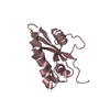
5
E: PR-1 protein
hetero molecules Summary Component details Symmetry operations Calculated values
Theoretical mass Number of molelcules Total (without water) 19,722 2 Polymers 19,599 1 Non-polymers 123 1 Water 0
Type Name Symmetry operation Number identity operation 1_555 x,y,z 1
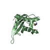
6
F: PR-1 protein
hetero molecules Summary Component details Symmetry operations Calculated values
Theoretical mass Number of molelcules Total (without water) 19,845 3 Polymers 19,599 1 Non-polymers 246 2 Water 0
Type Name Symmetry operation Number identity operation 1_555 x,y,z 1
7
G: PR-1 protein
hetero molecules Summary Component details Symmetry operations Calculated values
Theoretical mass Number of molelcules Total (without water) 19,968 4 Polymers 19,599 1 Non-polymers 369 3 Water 0
Type Name Symmetry operation Number identity operation 1_555 x,y,z 1
Unit cell Length a, b, c (Å) 108.412, 81.905, 130.238 Angle α, β, γ (deg.) 90.00, 111.64, 90.00 Int Tables number 4 Space group name H-M P121 1
Noncrystallographic symmetry (NCS) NCS domain Show large table (3 x 42) Hide large table ID Ens-ID Details (eV)1 1 A2 1 B1 2 A2 2 C1 3 A2 3 D1 4 A2 4 E1 5 A2 5 F1 6 A2 6 G1 7 B2 7 C1 8 B2 8 D1 9 B2 9 E1 10 B2 10 F1 11 B2 11 G1 12 C2 12 D1 13 C2 13 E1 14 C2 14 F1 15 C2 15 G1 16 D2 16 E1 17 D2 17 F1 18 D2 18 G1 19 E2 19 F1 20 E2 20 G1 21 F2 21 G
NCS domain segments Component-ID / Refine code
Show large table (10 x 42) Hide large table Dom-ID Ens-ID Beg auth comp-ID Beg label comp-ID End auth comp-ID End label comp-ID Auth asym-ID Label asym-ID Auth seq-ID Label seq-ID 1 1 PHEPHEGLYGLYAA32 - 163 10 - 141 2 1 PHEPHEGLYGLYBB32 - 163 10 - 141 1 2 PHEPHETYRTYRAA32 - 159 10 - 137 2 2 PHEPHETYRTYRCC32 - 159 10 - 137 1 3 PHEPHEASNASNAA32 - 161 10 - 139 2 3 PHEPHEASNASNDD32 - 161 10 - 139 1 4 PHEPHEGLYGLYAA32 - 163 10 - 141 2 4 PHEPHEGLYGLYEE32 - 163 10 - 141 1 5 PHEPHEALAALAAA32 - 162 10 - 140 2 5 PHEPHEALAALAFF32 - 162 10 - 140 1 6 PHEPHEGLYGLYAA32 - 163 10 - 141 2 6 PHEPHEGLYGLYGG32 - 163 10 - 141 1 7 PHEPHETYRTYRBB
 Movie
Movie Controller


 Open data
 Basic information
 Components
 Keywords
 Function and homology information
 Moniliophthora perniciosa (fungus)
 X-RAY DIFFRACTION /
  Authors
 Brazil, 1items
  Citation
 Structure visualization
 Downloads & links
 Other downloads

 PDBj
PDBj Assembly