Monochromator: SI 111 CHANNEL / Protocol: SINGLE WAVELENGTH / Monochromatic (M) / Laue (L): M / Scattering type: x-ray
Radiation wavelength
Wavelength: 0.9792 Å / Relative weight: 1
Reflection
Redundancy: 2.9 % / Av σ(I) over netI: 23.9 / Number: 210341 / Rmerge(I) obs: 0.046 / Χ2: 0.92 / D res high: 2.1 Å / D res low: 27 Å / Num. obs: 73135 / % possible obs: 98
Diffraction reflection shell
Highest resolution (Å)
Lowest resolution (Å)
% possible obs (%)
ID
Rmerge(I) obs
Chi squared
Redundancy
5.68
27
97.1
1
0.023
0.578
3.2
4.52
5.68
99.3
1
0.025
0.587
3.1
3.95
4.52
99
1
0.033
0.969
3.2
3.59
3.95
99.6
1
0.042
1.415
3.2
3.33
3.59
99.6
1
0.042
1.166
3.2
3.14
3.33
99.7
1
0.048
1.086
3.2
2.98
3.14
99.9
1
0.05
0.835
3.2
2.85
2.98
99.9
1
0.057
0.828
3.2
2.74
2.85
99.8
1
0.068
0.824
3.2
2.65
2.74
100
1
0.085
0.866
3.2
2.56
2.65
99.8
1
0.096
0.819
3.2
2.49
2.56
99.9
1
0.106
0.862
3.1
2.42
2.49
99.9
1
0.11
0.842
2.9
2.36
2.42
99.6
1
0.116
0.848
2.8
2.31
2.36
98.7
1
0.114
0.832
2.6
2.26
2.31
97.9
1
0.106
0.862
2.4
2.22
2.26
95.9
1
0.127
1.49
2.3
2.18
2.22
94.2
1
0.101
0.945
2.1
2.14
2.18
90.6
1
0.105
0.923
2
2.1
2.14
88.7
1
0.111
1.094
2
Reflection
Resolution: 2.1→50 Å / Num. obs: 73135 / % possible obs: 98 % / Redundancy: 2.9 % / Rmerge(I) obs: 0.046 / Χ2: 0.919 / Net I/σ(I): 13.8
Reflection shell
Resolution (Å)
Redundancy (%)
Rmerge(I) obs
Num. unique all
Χ2
Diffraction-ID
% possible all
2.1-2.14
2
0.111
3226
1.094
1
88.7
2.14-2.18
2
0.105
3477
0.923
1
90.6
2.18-2.22
2.1
0.101
3435
0.945
1
94.2
2.22-2.26
2.3
0.127
3680
1.49
1
95.9
2.26-2.31
2.4
0.106
3547
0.862
1
97.9
2.31-2.36
2.6
0.114
3687
0.832
1
98.7
2.36-2.42
2.8
0.116
3771
0.848
1
99.6
2.42-2.49
2.9
0.11
3693
0.842
1
99.9
2.49-2.56
3.1
0.106
3717
0.862
1
99.9
2.56-2.65
3.2
0.096
3741
0.819
1
99.8
2.65-2.74
3.2
0.085
3769
0.866
1
100
2.74-2.85
3.2
0.068
3674
0.824
1
99.8
2.85-2.98
3.2
0.057
3727
0.828
1
99.9
2.98-3.14
3.2
0.05
3746
0.835
1
99.9
3.14-3.33
3.2
0.048
3762
1.086
1
99.7
3.33-3.59
3.2
0.042
3649
1.166
1
99.6
3.59-3.95
3.2
0.042
3775
1.415
1
99.6
3.95-4.52
3.2
0.033
3720
0.969
1
99
4.52-5.68
3.1
0.025
3687
0.587
1
99.3
5.68-27
3.2
0.023
3652
0.578
1
97.1
-
Phasing
Phasing
Method: molecular replacement
-
Processing
Software
Name
Version
Classification
NB
DENZO
datareduction
SCALEPACK
datascaling
MOLREP
phasing
DM
phasing
REFMAC
refinement
PDB_EXTRACT
3.1
dataextraction
Refinement
Method to determine structure: MOLECULAR REPLACEMENT / Resolution: 2.1→27 Å / Cor.coef. Fo:Fc: 0.961 / Cor.coef. Fo:Fc free: 0.947 / Occupancy max: 1 / Occupancy min: 0.4 / SU B: 7.669 / SU ML: 0.108 / Cross valid method: THROUGHOUT / σ(F): 0 / ESU R Free: 0.174 / Stereochemistry target values: MAXIMUM LIKELIHOOD Details: U VALUES WITH TLS ADDED. HYDROGENS HAVE BEEN ADDED IN THE RIDING POSITIONS.
Rfactor
Num. reflection
% reflection
Selection details
Rfree
0.1911
3593
5 %
RANDOM
Rwork
0.157
-
-
-
obs
0.1587
71522
95.64 %
-
Solvent computation
Ion probe radii: 0.8 Å / Shrinkage radii: 0.8 Å / VDW probe radii: 1.2 Å / Solvent model: MASK
In the structure databanks used in Yorodumi, some data are registered as the other names, "COVID-19 virus" and "2019-nCoV". Here are the details of the virus and the list of structure data.
Jan 31, 2019. EMDB accession codes are about to change! (news from PDBe EMDB page)
EMDB accession codes are about to change! (news from PDBe EMDB page)
The allocation of 4 digits for EMDB accession codes will soon come to an end. Whilst these codes will remain in use, new EMDB accession codes will include an additional digit and will expand incrementally as the available range of codes is exhausted. The current 4-digit format prefixed with “EMD-” (i.e. EMD-XXXX) will advance to a 5-digit format (i.e. EMD-XXXXX), and so on. It is currently estimated that the 4-digit codes will be depleted around Spring 2019, at which point the 5-digit format will come into force.
The EM Navigator/Yorodumi systems omit the EMD- prefix.
Related info.:Q: What is EMD? / ID/Accession-code notation in Yorodumi/EM Navigator
Yorodumi is a browser for structure data from EMDB, PDB, SASBDB, etc.
This page is also the successor to EM Navigator detail page, and also detail information page/front-end page for Omokage search.
The word "yorodu" (or yorozu) is an old Japanese word meaning "ten thousand". "mi" (miru) is to see.
Related info.:EMDB / PDB / SASBDB / Comparison of 3 databanks / Yorodumi Search / Aug 31, 2016. New EM Navigator & Yorodumi / Yorodumi Papers / Jmol/JSmol / Function and homology information / Changes in new EM Navigator and Yorodumi
 Movie
Movie Controller

 Yorodumi
 Open data
 Basic information
 Components
 Keywords
 Function and homology information
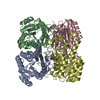
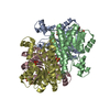
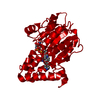
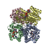
 Vibrio cholerae (bacteria)
 X-RAY DIFFRACTION /
  Authors
 Structure visualization
 Downloads & links
 Other downloads

 PDBj
PDBj Assembly

 Num. of mol.: 47 / Source method: obtained synthetically
 Mass: 96.063 Da / Num. of mol.: 19 / Source method: obtained synthetically / Formula: SO4
 Mass: 92.094 Da / Num. of mol.: 7 / Source method: obtained synthetically / Formula: C3H8O3
 Mass: 18.015 Da / Num. of mol.: 868 / Source method: isolated from a natural source / Formula: H2O
 Sample preparation
 / Beamline: 19-ID / Wavelength: 0.9792 Å
 Processing