[English] 日本語
Yorodumi
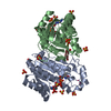
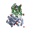
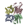
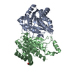
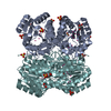
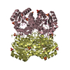
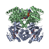
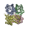
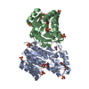
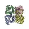
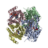
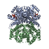
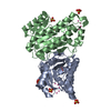
- PDB-3u09: Crystal structure of 3-ketoacyl-(acyl-carrier-protein) reductase ... -

+
Open data


ID or keywords:

Loading...

-
Basic information

Entry
Database: PDB / ID: 3u09
TitleCrystal structure of 3-ketoacyl-(acyl-carrier-protein) reductase (FabG)(G92D) from Vibrio cholerae
Components3-oxoacyl-[acyl-carrier-protein] reductase FabG
KeywordsOXIDOREDUCTASE / STRUCTURAL GENOMICS / CENTER FOR STRUCTURAL GENOMICS OF INFECTIOUS DISEASES / CSGID / Vibrio cholerae / 3-ketoacyl-(acyl-carrier-protein) reductase / FabG
Function / homology
Function and homology information


3-oxoacyl-[acyl-carrier-protein] reductase / 3-oxoacyl-[acyl-carrier-protein] reductase (NADPH) activity / fatty acid elongation / oxidoreductase activity, acting on the CH-OH group of donors, NAD or NADP as acceptor / NAD binding / NADP binding
Similarity search - Function
3-oxoacyl-(acyl-carrier-protein) reductase / PKS_KR / Short-chain dehydrogenase/reductase, conserved site / Short-chain dehydrogenases/reductases family signature. / Enoyl-(Acyl carrier protein) reductase / Short-chain dehydrogenase/reductase SDR / NAD(P)-binding Rossmann-like Domain / NAD(P)-binding domain superfamily / Rossmann fold / 3-Layer(aba) Sandwich / Alpha Beta
Similarity search - Domain/homology
3-oxoacyl-[acyl-carrier-protein] reductase FabG
Similarity search - Component
Biological speciesVibrio cholerae (bacteria)
MethodX-RAY DIFFRACTION / SYNCHROTRON / MOLECULAR REPLACEMENT / molecular replacement / Resolution: 1.75 Å
AuthorsHou, J. / Chruszcz, M. / Zheng, H. / Grabowski, M. / Fratczak, Z. / Anderson, W.F. / Minor, W. / Center for Structural Genomics of Infectious Diseases (CSGID)
CitationJournal: J.Bacteriol. / Year: 2015
Title: Dissecting the Structural Elements for the Activation of beta-Ketoacyl-(Acyl Carrier Protein) Reductase from Vibrio cholerae.
Authors: Hou, J. / Zheng, H. / Chruszcz, M. / Zimmerman, M.D. / Shumilin, I.A. / Osinski, T. / Demas, M. / Grimshaw, S. / Minor, W.
History
DepositionSep 28, 2011Deposition site: RCSB / Processing site: RCSB
Revision 1.0Oct 19, 2011Provider: repository / Type: Initial release
Revision 1.1Nov 12, 2014Group: Structure summary
Revision 1.2Nov 25, 2015Group: Database references
Revision 1.3Apr 6, 2016Group: Database references
Revision 1.4Aug 23, 2017Group: Data collection / Category: diffrn_detector / Item: _diffrn_detector.detector / _diffrn_detector.type
Revision 1.5Nov 8, 2017Group: Refinement description / Category: software
Item: _software.classification / _software.contact_author ..._software.classification / _software.contact_author / _software.contact_author_email / _software.date / _software.language / _software.location / _software.name / _software.type / _software.version
Revision 1.6Apr 13, 2022Group: Database references / Derived calculations / Structure summary
Category: audit_author / citation_author ...audit_author / citation_author / database_2 / struct_ref_seq_dif / struct_site
Item: _audit_author.identifier_ORCID / _citation_author.identifier_ORCID ..._audit_author.identifier_ORCID / _citation_author.identifier_ORCID / _database_2.pdbx_DOI / _database_2.pdbx_database_accession / _struct_ref_seq_dif.details / _struct_site.pdbx_auth_asym_id / _struct_site.pdbx_auth_comp_id / _struct_site.pdbx_auth_seq_id
Revision 1.7Feb 28, 2024Group: Data collection / Category: chem_comp_atom / chem_comp_bond

-
Structure visualization

Structure viewerMolecule:
MolmilJmol/JSmol

Downloads & links

-
Assembly

Deposited unit
A: 3-oxoacyl-[acyl-carrier-protein] reductase FabG
B: 3-oxoacyl-[acyl-carrier-protein] reductase FabG
hetero molecules


Theoretical massNumber of molelcules
Total (without water)53,60120
Polymers52,8322
Non-polymers76918
Water6,305350
1
A: 3-oxoacyl-[acyl-carrier-protein] reductase FabG
B: 3-oxoacyl-[acyl-carrier-protein] reductase FabG
hetero molecules

A: 3-oxoacyl-[acyl-carrier-protein] reductase FabG
B: 3-oxoacyl-[acyl-carrier-protein] reductase FabG
hetero molecules


Theoretical massNumber of molelcules
Total (without water)107,20240
Polymers105,6654
Non-polymers1,53736
Water724
TypeNameSymmetry operationNumber
identity operation1_555x,y,z1
crystal symmetry operation4_665-x+1,-y+1,z1
Buried area17110 Å2
ΔGint-280 kcal/mol
Surface area34370 Å2
MethodPISA
Unit cell
Length a, b, c (Å)63.574, 63.574, 191.059
Angle α, β, γ (deg.)90.000, 90.000, 120.000
Int Tables number171
Space group name H-MP62
Components on special symmetry positions
IDModelComponents
11A-347-

HOH

-
Components

#1: Protein 3-oxoacyl-[acyl-carrier-protein] reductase FabG / 3-ketoacyl-acyl carrier protein reductase / Beta-Ketoacyl-acyl carrier protein reductase / Beta- ...3-ketoacyl-acyl carrier protein reductase / Beta-Ketoacyl-acyl carrier protein reductase / Beta-ketoacyl-ACP reductase


Mass: 26416.148 Da / Num. of mol.: 2 / Mutation: G92D
Source method: isolated from a genetically manipulated source
Source: (gene. exp.) Vibrio cholerae (bacteria) / Strain: O1 biovar eltor str. N16961 / Gene: fabG, VC2021, VC_2021 / Plasmid: pMCSG7 / Production host: Escherichia coli (E. coli) / Strain (production host): BL21-CodonPlus(DE3)-RIPL
References: UniProt: Q9KQH7, 3-oxoacyl-[acyl-carrier-protein] reductase
#2: Chemical
ChemComp-SO4 / SULFATE ION / Sulfate


Mass: 96.063 Da / Num. of mol.: 8 / Source method: obtained synthetically / Formula: SO4
#3: Chemical
ChemComp-UNX / UNKNOWN ATOM OR ION


Num. of mol.: 10 / Source method: obtained synthetically
#4: Water ChemComp-HOH / water / Water


Mass: 18.015 Da / Num. of mol.: 350 / Source method: isolated from a natural source / Formula: H2O

-
Experimental details

-
Experiment

ExperimentMethod: X-RAY DIFFRACTION / Number of used crystals: 1

-
Sample preparation

CrystalDensity Matthews: 2.11 Å3/Da / Density % sol: 41.7 %
Crystal growMethod: vapor diffusion / pH: 9
Details: 0.1M Tris, 2.3M Ammonium Sulfate, 5%Tacsimate, pH 9, vapor diffusion

-
Data collection

DiffractionMean temperature: 100 K
Diffraction sourceSource: SYNCHROTRON / Site: APS / Beamline: 19-BM / Wavelength: 0.9792 Å
DetectorType: ADSC QUANTUM 210 / Detector: CCD / Date: Jul 6, 2010 / Details: MIRRORS
RadiationMonochromator: SI 111 CHANNEL / Protocol: SINGLE WAVELENGTH / Monochromatic (M) / Laue (L): M / Scattering type: x-ray
Radiation wavelengthWavelength: 0.9792 Å / Relative weight: 1
ReflectionRedundancy: 4.6 % / Av σ(I) over netI: 38.05 / Number: 194251 / Rmerge(I) obs: 0.054 / Χ2: 2.03 / D res high: 1.75 Å / D res low: 27 Å / Num. obs: 42687 / % possible obs: 97.2
Diffraction reflection shell
Highest resolution (Å)Lowest resolution (Å)% possible obs (%)IDRmerge(I) obsChi squaredRedundancy
4.742799.310.0413.0936.1
3.774.7499.910.0392.8066.3
3.293.7799.710.0452.9686.3
2.993.2910010.052.8586
2.782.9999.910.0552.6125.4
2.612.7899.810.0622.4234.8
2.482.6199.710.0682.434.3
2.372.4898.710.0762.274
2.282.3798.510.082.023.8
2.22.2896.810.0922.0193.8
2.142.295.710.0971.8173.7
2.072.1494.710.1131.6423.8
2.022.0794.710.121.3623.9
1.972.0294.710.1531.2374
1.931.9795.310.1951.2124
1.891.9396.110.2351.24
1.851.8995.710.2770.9484.1
1.811.8594.610.3340.8574.2
1.781.8195.310.3610.8224.1
1.751.7895.810.420.7234.1
Reflection twin
Crystal-IDIDOperatorDomain-IDFraction
11H, K, L10.833
11K, H, -L20.167
ReflectionResolution: 1.75→27 Å / Num. obs: 42687 / % possible obs: 97.2 % / Redundancy: 4.6 % / Rmerge(I) obs: 0.054 / Χ2: 2.033 / Net I/σ(I): 20.1
Reflection shell
Resolution (Å)Redundancy (%)Rmerge(I) obsNum. unique allΧ2Diffraction-ID% possible all
1.75-1.784.10.4220780.723195.8
1.78-1.814.10.36120980.822195.3
1.81-1.854.20.33420700.857194.6
1.85-1.894.10.27720740.948195.7
1.89-1.9340.23521451.2196.1
1.93-1.9740.19520561.212195.3
1.97-2.0240.15320531.237194.7
2.02-2.073.90.1220911.362194.7
2.07-2.143.80.11320711.642194.7
2.14-2.23.70.09721251.817195.7
2.2-2.283.80.09220912.019196.8
2.28-2.373.80.0821602.02198.5
2.37-2.4840.07621792.27198.7
2.48-2.614.30.06821852.43199.7
2.61-2.784.80.06221832.423199.8
2.78-2.995.40.05522002.612199.9
2.99-3.2960.0521872.8581100
3.29-3.776.30.04522052.968199.7
3.77-4.746.30.03922062.806199.9
4.74-276.10.04122303.093199.3

-
Phasing

PhasingMethod: molecular replacement

-
Processing

Software
NameVersionClassificationNB
DENZOdata reduction
SCALEPACKdata scaling
MOLREPphasing
DMphasing
REFMACrefinement
PDB_EXTRACT3.1data extraction
RefinementMethod to determine structure: MOLECULAR REPLACEMENT / Resolution: 1.75→27 Å / Cor.coef. Fo:Fc: 0.966 / Cor.coef. Fo:Fc free: 0.954 / Occupancy max: 1 / Occupancy min: 0.2 / SU B: 3.859 / SU ML: 0.062 / Cross valid method: THROUGHOUT / σ(F): 0 / ESU R Free: 0.023 / Stereochemistry target values: MAXIMUM LIKELIHOOD
Details: U VALUES : WITH TLS ADDED HYDROGENS HAVE BEEN ADDED IN THE RIDING POSITIONS
RfactorNum. reflection% reflectionSelection details
Rfree0.187 2259 5.3 %RANDOM
Rwork0.1598 ---
obs0.1613 42619 97.09 %-
Solvent computationIon probe radii: 0.8 Å / Shrinkage radii: 0.8 Å / VDW probe radii: 1.2 Å / Solvent model: MASK
Displacement parametersBiso max: 69.83 Å2 / Biso mean: 27.0196 Å2 / Biso min: 13.32 Å2
Baniso -1Baniso -2Baniso -3
1--1.85 Å20 Å20 Å2
2---1.85 Å20 Å2
3---3.7 Å2
Refinement stepCycle: LAST / Resolution: 1.75→27 Å
ProteinNucleic acidLigandSolventTotal
Num. atoms3613 0 50 350 4013
Refine LS restraints
Refine-IDTypeDev idealDev ideal targetNumber
X-RAY DIFFRACTIONr_bond_refined_d0.010.0193711
X-RAY DIFFRACTIONr_bond_other_d0.0030.022434
X-RAY DIFFRACTIONr_angle_refined_deg1.3081.9755019
X-RAY DIFFRACTIONr_angle_other_deg1.02735973
X-RAY DIFFRACTIONr_dihedral_angle_1_deg5.3515502
X-RAY DIFFRACTIONr_dihedral_angle_2_deg36.56924.752141
X-RAY DIFFRACTIONr_dihedral_angle_3_deg12.39115644
X-RAY DIFFRACTIONr_dihedral_angle_4_deg16.0871524
X-RAY DIFFRACTIONr_chiral_restr0.0730.2597
X-RAY DIFFRACTIONr_gen_planes_refined0.0060.024182
X-RAY DIFFRACTIONr_gen_planes_other0.0030.02702
LS refinement shellResolution: 1.749→1.795 Å / Total num. of bins used: 20
RfactorNum. reflection% reflection
Rfree0.247 140 -
Rwork0.23 2861 -
all-3001 -
obs--93.69 %
Refinement TLS params.Method: refined / Origin x: 2.654 Å / Origin y: 19.766 Å / Origin z: -0.099 Å
111213212223313233
T0.132 Å2-0.0407 Å20.02 Å2-0.0751 Å2-0.0211 Å2--0.0163 Å2
L0.7303 °20.4121 °2-0.0628 °2-0.4501 °2-0.0521 °2--0.5265 °2
S0.0861 Å °0.1169 Å °-0.0095 Å °0.1685 Å °-0.0368 Å °0.0458 Å °0.0509 Å °-0.054 Å °-0.0493 Å °
Refinement TLS group
IDRefine-IDRefine TLS-IDAuth asym-IDAuth seq-ID
1X-RAY DIFFRACTION1A2 - 248
2X-RAY DIFFRACTION1B2 - 248

+
About Yorodumi

-
News

-
Feb 9, 2022. New format data for meta-information of EMDB entries

New format data for meta-information of EMDB entries

  • Version 3 of the EMDB header file is now the official format.
  • The previous official version 1.9 will be removed from the archive.

Related info.:EMDB header

External links:wwPDB to switch to version 3 of the EMDB data model

-
Aug 12, 2020. Covid-19 info

Covid-19 info

URL: https://pdbj.org/emnavi/covid19.php

New page: Covid-19 featured information page in EM Navigator.

Related info.:Covid-19 info / Mar 5, 2020. Novel coronavirus structure data

+
Mar 5, 2020. Novel coronavirus structure data

Novel coronavirus structure data

Related info.:Yorodumi Speices / Aug 12, 2020. Covid-19 info

External links:COVID-19 featured content - PDBj / Molecule of the Month (242):Coronavirus Proteases

+
Jan 31, 2019. EMDB accession codes are about to change! (news from PDBe EMDB page)

EMDB accession codes are about to change! (news from PDBe EMDB page)

  • The allocation of 4 digits for EMDB accession codes will soon come to an end. Whilst these codes will remain in use, new EMDB accession codes will include an additional digit and will expand incrementally as the available range of codes is exhausted. The current 4-digit format prefixed with “EMD-” (i.e. EMD-XXXX) will advance to a 5-digit format (i.e. EMD-XXXXX), and so on. It is currently estimated that the 4-digit codes will be depleted around Spring 2019, at which point the 5-digit format will come into force.
  • The EM Navigator/Yorodumi systems omit the EMD- prefix.

Related info.:Q: What is EMD? / ID/Accession-code notation in Yorodumi/EM Navigator

External links:EMDB Accession Codes are Changing Soon! / Contact to PDBj

+
Jul 12, 2017. Major update of PDB

Major update of PDB

  • wwPDB released updated PDB data conforming to the new PDBx/mmCIF dictionary.
  • This is a major update changing the version number from 4 to 5, and with Remediation, in which all the entries are updated.
  • In this update, many items about electron microscopy experimental information are reorganized (e.g. em_software).
  • Now, EM Navigator and Yorodumi are based on the updated data.

External links:wwPDB Remediation / Enriched Model Files Conforming to OneDep Data Standards Now Available in the PDB FTP Archive

-
Yorodumi

Thousand views of thousand structures

  • Yorodumi is a browser for structure data from EMDB, PDB, SASBDB, etc.
  • This page is also the successor to EM Navigator detail page, and also detail information page/front-end page for Omokage search.
  • The word "yorodu" (or yorozu) is an old Japanese word meaning "ten thousand". "mi" (miru) is to see.

Related info.:EMDB / PDB / SASBDB / Comparison of 3 databanks / Yorodumi Search / Aug 31, 2016. New EM Navigator & Yorodumi / Yorodumi Papers / Jmol/JSmol / Function and homology information / Changes in new EM Navigator and Yorodumi

Read more