| Entry | Database: PDB / ID: 6k79
|
|---|
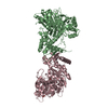
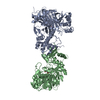
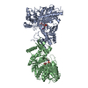
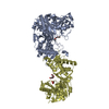
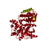
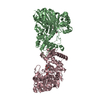
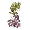
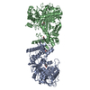
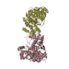
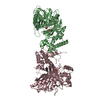
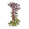
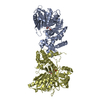
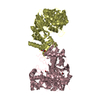
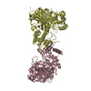
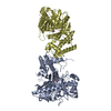
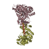
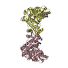
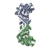
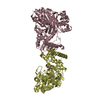
| Title | Glycerol kinase form Thermococcus kodakarensis, complex structure with substrate. |
|---|
 Components Components | (Glycerol kinase) x 2 |
|---|
 Keywords Keywords | TRANSFERASE / Glycerol / Kinase / ATP / Archaea |
|---|
| Function / homology |  Function and homology information Function and homology information
glycerol-3-phosphate metabolic process / glycerol kinase / glycerol kinase activity / glycerol metabolic process / glycerol catabolic process / ATP binding / cytosolSimilarity search - Function FGGY family of carbohydrate kinases signature 1. / Glycerol kinase / FGGY family of carbohydrate kinases signature 2. / Carbohydrate kinase, FGGY, conserved site / Carbohydrate kinase, FGGY / Carbohydrate kinase, FGGY, N-terminal / FGGY family of carbohydrate kinases, N-terminal domain / Carbohydrate kinase, FGGY, C-terminal / FGGY family of carbohydrate kinases, C-terminal domain / ATPase, nucleotide binding domain ...FGGY family of carbohydrate kinases signature 1. / Glycerol kinase / FGGY family of carbohydrate kinases signature 2. / Carbohydrate kinase, FGGY, conserved site / Carbohydrate kinase, FGGY / Carbohydrate kinase, FGGY, N-terminal / FGGY family of carbohydrate kinases, N-terminal domain / Carbohydrate kinase, FGGY, C-terminal / FGGY family of carbohydrate kinases, C-terminal domain / ATPase, nucleotide binding domain / ATPase, nucleotide binding domain / Nucleotidyltransferase; domain 5 / 2-Layer Sandwich / Alpha BetaSimilarity search - Domain/homology |
|---|
| Biological species |   Thermococcus kodakarensis (archaea) Thermococcus kodakarensis (archaea) |
|---|
| Method |  X-RAY DIFFRACTION / SYNCHROTRON / MOLECULAR REPLACEMENT / Resolution: 2.19 Å X-RAY DIFFRACTION / SYNCHROTRON / MOLECULAR REPLACEMENT / Resolution: 2.19 Å |
|---|
 Authors Authors | Koga, Y. / Angkawidjaja, C. / Matsumura, H. / Hokao, R. |
|---|
| Funding support |  Japan, 1items Japan, 1items | Organization | Grant number | Country |
|---|
| Ministry of Education, Culture, Sports, Science and Technology (Japan) | JP22780091 | Japan |
|
|---|
 Citation Citation | Journal: To Be Published
Title: Structural analysis of hexameric structure of glycerol kinase from Thermococcus kodakaraeinsis KOD1
Authors: Hokao, R. / Matsumura, H. / Katsumi, R. / Angkawidjaja, C. / Takano, K. / Kanaya, S. / Koga, Y. |
|---|
| History | | Deposition | Jun 6, 2019 | Deposition site: PDBJ / Processing site: PDBJ |
|---|
| Revision 1.0 | Jun 10, 2020 | Provider: repository / Type: Initial release |
|---|
| Revision 1.1 | Mar 27, 2024 | Group: Data collection / Database references / Category: chem_comp_atom / chem_comp_bond / database_2
Item: _database_2.pdbx_DOI / _database_2.pdbx_database_accession |
|---|
|
|---|
 Movie
Movie Controller

 Yorodumi
 Open data
 Basic information
 Structure visualization
 Downloads & links
 Other downloads
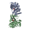


 PDBj
PDBj Assembly


 Mass: 150.173 Da / Num. of mol.: 4 / Source method: obtained synthetically / Formula: C6H14O4
 Mass: 92.094 Da / Num. of mol.: 4 / Source method: obtained synthetically / Formula: C3H8O3
 Mass: 18.015 Da / Num. of mol.: 457 / Source method: isolated from a natural source / Formula: H2O
 Sample preparation
 Processing