 Movie
Movie Controller

[English] 日本語
 Yorodumi
- PDB-6zq8: Crystal structure of Chaetomium thermophilum Glycerol Kinase in P... -
+ Open data

- Basic information
| Entry | Database: PDB / ID: 6zq8 | |||||||||
|---|---|---|---|---|---|---|---|---|---|---|
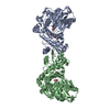
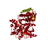
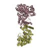
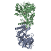
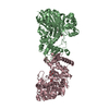
| Title | Crystal structure of Chaetomium thermophilum Glycerol Kinase in P3221 space group | |||||||||
 Components Components | Glycerol kinase-like protein | |||||||||
 Keywords Keywords | TRANSFERASE / Kinase / Glycerol / Metabolism | |||||||||
| Function / homology |  Function and homology information Function and homology informationglycerol-3-phosphate biosynthetic process / glycerol kinase / glycerol kinase activity / glycerol catabolic process / triglyceride metabolic process / mitochondrion / ATP binding Similarity search - Function | |||||||||
| Biological species |  Chaetomium thermophilum (fungus) Chaetomium thermophilum (fungus) | |||||||||
| Method |  X-RAY DIFFRACTION / SYNCHROTRON / MOLECULAR REPLACEMENT / Resolution: 2.38 Å X-RAY DIFFRACTION / SYNCHROTRON / MOLECULAR REPLACEMENT / Resolution: 2.38 Å | |||||||||
 Authors Authors | Wilk, P. / Wator, E. / Malecki, P. / Tokarz, P. / Grudnik, P. | |||||||||
| Funding support |  Poland, 2items Poland, 2items
| |||||||||
 Citation Citation | Journal: Int J Mol Sci / Year: 2020 Title: Structural Characterization of Glycerol Kinase from the Thermophilic Fungus Chaetomium thermophilum . Authors: Wilk, P. / Kuska, K. / Wator, E. / Malecki, P.H. / Wos, K. / Tokarz, P. / Dubin, G. / Grudnik, P. | |||||||||
| History |
|
- Structure visualization
| Structure viewer | Molecule: MolmilJmol/JSmol |
|---|
- Downloads & links
-Download
| PDBx/mmCIF format | 6zq8.cif.gz | 572.8 KB | Display | PDBx/mmCIF format |
|---|---|---|---|---|
| PDB format | pdb6zq8.ent.gz | 481.8 KB | Display | PDB format |
| PDBx/mmJSON format | 6zq8.json.gz | Tree view | PDBx/mmJSON format | |
| Others |  Other downloads Other downloads |
-Validation report
| Arichive directory | https://data.pdbj.org/pub/pdb/validation_reports/zq/6zq8ftp://data.pdbj.org/pub/pdb/validation_reports/zq/6zq8 | HTTPS FTP |
|---|
-Related structure data
| Related structure data | 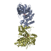 6zq4C 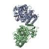 6zq5C 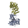 6zq6C  6zq7C  2d4wS S: Starting model for refinement C: citing same article ( |
|---|---|
| Similar structure data |


-Links
 PDBj
PDBj- Assembly
| Deposited unit | 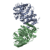
| ||||||||
|---|---|---|---|---|---|---|---|---|---|
| 1 | 
| ||||||||
| Unit cell |
| ||||||||
| Components on special symmetry positions |
|
-Components
| #1: Protein | Mass: 57067.277 Da / Num. of mol.: 2 Source method: isolated from a genetically manipulated source Source: (gene. exp.) Chaetomium thermophilum (strain DSM 1495 / CBS 144.50 / IMI 039719) (fungus)Gene: CTHT_0042250 / Production host:  #2: Water | ChemComp-HOH / |  Mass: 18.015 Da / Num. of mol.: 74 / Source method: isolated from a natural source / Formula: H2O Mass: 18.015 Da / Num. of mol.: 74 / Source method: isolated from a natural source / Formula: H2O |
|---|
-Experimental details
-Experiment
| Experiment | Method: X-RAY DIFFRACTION / Number of used crystals: 1 |
|---|
- Sample preparation
| Crystal | Density Matthews: 2.5 Å3/Da / Density % sol: 50.83 % |
|---|---|
| Crystal grow | Temperature: 293 K / Method: vapor diffusion, sitting drop / Details: 0.125-0.3 M CaCl2; 16-19% PEG 3350 |
-Data collection
| Diffraction | Mean temperature: 100 K / Serial crystal experiment: N | ||||||||||||||||||||||||||||||||||||||||||||||||||||||||||||||||||||||||||||||||||||||||||||||||||||
|---|---|---|---|---|---|---|---|---|---|---|---|---|---|---|---|---|---|---|---|---|---|---|---|---|---|---|---|---|---|---|---|---|---|---|---|---|---|---|---|---|---|---|---|---|---|---|---|---|---|---|---|---|---|---|---|---|---|---|---|---|---|---|---|---|---|---|---|---|---|---|---|---|---|---|---|---|---|---|---|---|---|---|---|---|---|---|---|---|---|---|---|---|---|---|---|---|---|---|---|---|---|
| Diffraction source | Source: SYNCHROTRON / Site: PETRA III, DESY  / Beamline: P11 / Wavelength: 1.033 Å / Beamline: P11 / Wavelength: 1.033 Å | ||||||||||||||||||||||||||||||||||||||||||||||||||||||||||||||||||||||||||||||||||||||||||||||||||||
| Detector | Type: DECTRIS PILATUS 6M / Detector: PIXEL / Date: May 27, 2018 | ||||||||||||||||||||||||||||||||||||||||||||||||||||||||||||||||||||||||||||||||||||||||||||||||||||
| Radiation | Protocol: SINGLE WAVELENGTH / Monochromatic (M) / Laue (L): M / Scattering type: x-ray | ||||||||||||||||||||||||||||||||||||||||||||||||||||||||||||||||||||||||||||||||||||||||||||||||||||
| Radiation wavelength | Wavelength: 1.033 Å / Relative weight: 1 | ||||||||||||||||||||||||||||||||||||||||||||||||||||||||||||||||||||||||||||||||||||||||||||||||||||
| Reflection | Resolution: 2.38→47.04 Å / Num. obs: 46200 / % possible obs: 98.8 % / Redundancy: 19.242 % / Biso Wilson estimate: 66.893 Å2 / CC1/2: 0.999 / Rmerge(I) obs: 0.124 / Rrim(I) all: 0.127 / Χ2: 1.077 / Net I/σ(I): 17.95 / Num. measured all: 888970 | ||||||||||||||||||||||||||||||||||||||||||||||||||||||||||||||||||||||||||||||||||||||||||||||||||||
| Reflection shell | Diffraction-ID: 1
|
- Processing
| Software |
| ||||||||||||||||||||||||||||||||||||||||||||||||||||||||||||||||||||||||||||||||||||||||||||||||||||||||||||||||||||||||||||||||||||||||||||||||||||||
|---|---|---|---|---|---|---|---|---|---|---|---|---|---|---|---|---|---|---|---|---|---|---|---|---|---|---|---|---|---|---|---|---|---|---|---|---|---|---|---|---|---|---|---|---|---|---|---|---|---|---|---|---|---|---|---|---|---|---|---|---|---|---|---|---|---|---|---|---|---|---|---|---|---|---|---|---|---|---|---|---|---|---|---|---|---|---|---|---|---|---|---|---|---|---|---|---|---|---|---|---|---|---|---|---|---|---|---|---|---|---|---|---|---|---|---|---|---|---|---|---|---|---|---|---|---|---|---|---|---|---|---|---|---|---|---|---|---|---|---|---|---|---|---|---|---|---|---|---|---|---|---|
| Refinement | Method to determine structure: MOLECULAR REPLACEMENT Starting model: 2d4w Resolution: 2.38→47.04 Å / SU ML: 0.4 / Cross valid method: THROUGHOUT / σ(F): 1.33 / Phase error: 32.55 / Stereochemistry target values: ML
| ||||||||||||||||||||||||||||||||||||||||||||||||||||||||||||||||||||||||||||||||||||||||||||||||||||||||||||||||||||||||||||||||||||||||||||||||||||||
| Solvent computation | Shrinkage radii: 0.9 Å / VDW probe radii: 1.11 Å / Solvent model: FLAT BULK SOLVENT MODEL | ||||||||||||||||||||||||||||||||||||||||||||||||||||||||||||||||||||||||||||||||||||||||||||||||||||||||||||||||||||||||||||||||||||||||||||||||||||||
| Displacement parameters | Biso max: 222.54 Å2 / Biso mean: 88.2821 Å2 / Biso min: 36.25 Å2 | ||||||||||||||||||||||||||||||||||||||||||||||||||||||||||||||||||||||||||||||||||||||||||||||||||||||||||||||||||||||||||||||||||||||||||||||||||||||
| Refinement step | Cycle: final / Resolution: 2.38→47.04 Å
| ||||||||||||||||||||||||||||||||||||||||||||||||||||||||||||||||||||||||||||||||||||||||||||||||||||||||||||||||||||||||||||||||||||||||||||||||||||||
| LS refinement shell | Refine-ID: X-RAY DIFFRACTION / Rfactor Rfree error: 0 / Total num. of bins used: 15
| ||||||||||||||||||||||||||||||||||||||||||||||||||||||||||||||||||||||||||||||||||||||||||||||||||||||||||||||||||||||||||||||||||||||||||||||||||||||
| Refinement TLS params. | Method: refined / Refine-ID: X-RAY DIFFRACTION
| ||||||||||||||||||||||||||||||||||||||||||||||||||||||||||||||||||||||||||||||||||||||||||||||||||||||||||||||||||||||||||||||||||||||||||||||||||||||
| Refinement TLS group |
|
