 Movie
Movie Controller

[English] 日本語
 Yorodumi
- PDB-6j9q: Crystal structure of Trypanosoma brucei gambiense glycerol kinase... -
+ Open data

- Basic information
| Entry | Database: PDB / ID: 6j9q | ||||||
|---|---|---|---|---|---|---|---|
| Title | Crystal structure of Trypanosoma brucei gambiense glycerol kinase complex with AMP-PNP. | ||||||
 Components Components | Glycerol kinase | ||||||
 Keywords Keywords | TRANSFERASE / TRYPANOSOMA / GLYCEROL KINASE / SUGAR KINASE SUPERFAMILY / GLYCOSOME | ||||||
| Function / homology |  Function and homology information Function and homology informationglycerol-3-phosphate biosynthetic process / glycerol kinase / glycerol kinase activity / glycerol catabolic process / triglyceride metabolic process / mitochondrion / ATP binding Similarity search - Function | ||||||
| Biological species |  | ||||||
| Method |  X-RAY DIFFRACTION / SYNCHROTRON / MOLECULAR REPLACEMENT / Resolution: 2.7 Å X-RAY DIFFRACTION / SYNCHROTRON / MOLECULAR REPLACEMENT / Resolution: 2.7 Å | ||||||
 Authors Authors | Balogun, E.O. / Chishima, T. / Ichinose, M. / Inaoka, D.K. / Kido, Y. / Ibrahim, B. / de Koning, H. / McKerrow, J.H. / Watanabe, Y. / Nozaki, T. ...Balogun, E.O. / Chishima, T. / Ichinose, M. / Inaoka, D.K. / Kido, Y. / Ibrahim, B. / de Koning, H. / McKerrow, J.H. / Watanabe, Y. / Nozaki, T. / Michels, P.A.M. / Harada, S. / Kita, K. / Shiba, T. | ||||||
 Citation Citation | Journal: To Be Published Title: Reaction mechanism of the reverse reaction of African human trypanosomes glycerol kinase. Authors: Balogun, E.O. / Chishima, T. / Ichinose, M. / Inaoka, D.K. / Kido, Y. / Ibrahim, B. / de Koning, H. / McKerrow, J.H. / Watanabe, Y. / Nozaki, T. / Michels, P.A.M. / Harada, S. / Kita, K. / Shiba, T. | ||||||
| History |
|
- Structure visualization
| Structure viewer | Molecule: MolmilJmol/JSmol |
|---|
- Downloads & links
-Download
| PDBx/mmCIF format | 6j9q.cif.gz | 399.4 KB | Display | PDBx/mmCIF format |
|---|---|---|---|---|
| PDB format | pdb6j9q.ent.gz | 325.9 KB | Display | PDB format |
| PDBx/mmJSON format | 6j9q.json.gz | Tree view | PDBx/mmJSON format | |
| Others |  Other downloads Other downloads |
-Validation report
| Arichive directory | https://data.pdbj.org/pub/pdb/validation_reports/j9/6j9qftp://data.pdbj.org/pub/pdb/validation_reports/j9/6j9q | HTTPS FTP |
|---|
-Related structure data
| Related structure data |  6j9vC  3wxlS S: Starting model for refinement C: citing same article ( |
|---|---|
| Similar structure data |


-Links
 PDBj
PDBj


- Assembly
| Deposited unit | 
| ||||||||
|---|---|---|---|---|---|---|---|---|---|
| 1 | 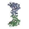
| ||||||||
| 2 | 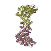
| ||||||||
| Unit cell |
|
-Components
| #1: Protein | Mass: 57064.625 Da / Num. of mol.: 4 Source method: isolated from a genetically manipulated source Source: (gene. exp.) Gene: gk / Plasmid: PET151/D-TOPO / Production host:  #2: Chemical | ChemComp-GOL /   Mass: 92.094 Da / Num. of mol.: 6 / Source method: obtained synthetically / Formula: C3H8O3 Mass: 92.094 Da / Num. of mol.: 6 / Source method: obtained synthetically / Formula: C3H8O3#3: Chemical | ChemComp-ANP /   Mass: 506.196 Da / Num. of mol.: 4 / Source method: obtained synthetically / Formula: C10H17N6O12P3 / Comment: AMP-PNP, energy-carrying molecule analogue*YM Mass: 506.196 Da / Num. of mol.: 4 / Source method: obtained synthetically / Formula: C10H17N6O12P3 / Comment: AMP-PNP, energy-carrying molecule analogue*YM#4: Water | ChemComp-HOH / |  Mass: 18.015 Da / Num. of mol.: 19 / Source method: isolated from a natural source / Formula: H2O Mass: 18.015 Da / Num. of mol.: 19 / Source method: isolated from a natural source / Formula: H2OHas protein modification | Y | |
|---|
-Experimental details
-Experiment
| Experiment | Method: X-RAY DIFFRACTION / Number of used crystals: 1 |
|---|
- Sample preparation
| Crystal | Density Matthews: 2.6 Å3/Da / Density % sol: 52.77 % |
|---|---|
| Crystal grow | Temperature: 293 K / Method: vapor diffusion, hanging drop / pH: 7.5 / Details: 8-18% PEG 400, 0.1M HEPES, 11% 1,6-HEXANEDIOL |
-Data collection
| Diffraction | Mean temperature: 100 K / Serial crystal experiment: N |
|---|---|
| Diffraction source | Source: SYNCHROTRON / Site: Photon Factory  / Beamline: BL-17A / Wavelength: 0.98 Å / Beamline: BL-17A / Wavelength: 0.98 Å |
| Detector | Type: DECTRIS EIGER X 16M / Detector: PIXEL / Date: Nov 30, 2018 |
| Radiation | Monochromator: SI(111) / Protocol: SINGLE WAVELENGTH / Monochromatic (M) / Laue (L): M / Scattering type: x-ray |
| Radiation wavelength | Wavelength: 0.98 Å / Relative weight: 1 |
| Reflection | Resolution: 2.7→50 Å / Num. obs: 62097 / % possible obs: 98.4 % / Redundancy: 2.7 % / CC1/2: 0.998 / Rmerge(I) obs: 0.054 / Rpim(I) all: 0.068 / Net I/σ(I): 12.5 |
| Reflection shell | Resolution: 2.7→2.86 Å / Redundancy: 2.7 % / Rmerge(I) obs: 0.448 / Mean I/σ(I) obs: 2 / Num. unique obs: 20049 / % possible all: 98 |
- Processing
| Software |
| ||||||||||||||||||||||||||||||||||||||||||||||||||||||||||||||||||||||||||||||||||||||||||||||||||||||||||||||||||||||||||||||||||||||||||||||||||||||||||||||||||||||||||||||||||||||
|---|---|---|---|---|---|---|---|---|---|---|---|---|---|---|---|---|---|---|---|---|---|---|---|---|---|---|---|---|---|---|---|---|---|---|---|---|---|---|---|---|---|---|---|---|---|---|---|---|---|---|---|---|---|---|---|---|---|---|---|---|---|---|---|---|---|---|---|---|---|---|---|---|---|---|---|---|---|---|---|---|---|---|---|---|---|---|---|---|---|---|---|---|---|---|---|---|---|---|---|---|---|---|---|---|---|---|---|---|---|---|---|---|---|---|---|---|---|---|---|---|---|---|---|---|---|---|---|---|---|---|---|---|---|---|---|---|---|---|---|---|---|---|---|---|---|---|---|---|---|---|---|---|---|---|---|---|---|---|---|---|---|---|---|---|---|---|---|---|---|---|---|---|---|---|---|---|---|---|---|---|---|---|---|
| Refinement | Method to determine structure: MOLECULAR REPLACEMENT Starting model: 3WXL Resolution: 2.7→19.88 Å / Cor.coef. Fo:Fc: 0.956 / Cor.coef. Fo:Fc free: 0.91 / SU B: 15.084 / SU ML: 0.306 / Cross valid method: THROUGHOUT / ESU R Free: 0.38 / Stereochemistry target values: MAXIMUM LIKELIHOOD / Details: HYDROGENS HAVE BEEN ADDED IN THE RIDING POSITIONS
| ||||||||||||||||||||||||||||||||||||||||||||||||||||||||||||||||||||||||||||||||||||||||||||||||||||||||||||||||||||||||||||||||||||||||||||||||||||||||||||||||||||||||||||||||||||||
| Solvent computation | Ion probe radii: 0.8 Å / Shrinkage radii: 0.8 Å / VDW probe radii: 1.2 Å / Solvent model: MASK | ||||||||||||||||||||||||||||||||||||||||||||||||||||||||||||||||||||||||||||||||||||||||||||||||||||||||||||||||||||||||||||||||||||||||||||||||||||||||||||||||||||||||||||||||||||||
| Displacement parameters | Biso mean: 70.957 Å2
| ||||||||||||||||||||||||||||||||||||||||||||||||||||||||||||||||||||||||||||||||||||||||||||||||||||||||||||||||||||||||||||||||||||||||||||||||||||||||||||||||||||||||||||||||||||||
| Refinement step | Cycle: 1 / Resolution: 2.7→19.88 Å
| ||||||||||||||||||||||||||||||||||||||||||||||||||||||||||||||||||||||||||||||||||||||||||||||||||||||||||||||||||||||||||||||||||||||||||||||||||||||||||||||||||||||||||||||||||||||
| Refine LS restraints |
|