 Movie
Movie Controller

[English] 日本語
 Yorodumi
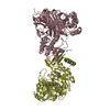
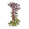
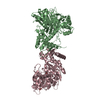
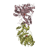
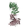
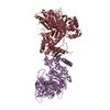
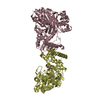
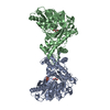
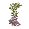
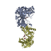
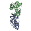
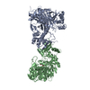
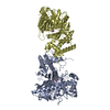
- PDB-2d4w: Crystal structure of glycerol kinase from Cellulomonas sp. NT3060 -
+ Open data

- Basic information
| Entry | Database: PDB / ID: 2d4w | ||||||
|---|---|---|---|---|---|---|---|
| Title | Crystal structure of glycerol kinase from Cellulomonas sp. NT3060 | ||||||
 Components Components | glycerol kinase | ||||||
 Keywords Keywords | TRANSFERASE / Alpha and beta protein / Ribonuclease H-like motif / Actin-like ATPase domain | ||||||
| Function / homology | ATPase, nucleotide binding domain / Nucleotidyltransferase; domain 5 / 2-Layer Sandwich / Alpha Beta Function and homology information Function and homology information | ||||||
| Biological species |  Cellulomonas sp. (bacteria) Cellulomonas sp. (bacteria) | ||||||
| Method |  X-RAY DIFFRACTION / SYNCHROTRON / SIROAS / Resolution: 2.3 Å X-RAY DIFFRACTION / SYNCHROTRON / SIROAS / Resolution: 2.3 Å | ||||||
 Authors Authors | Imada, K. / Tamura, T. / Namba, K. / Inagaki, K. | ||||||
 Citation Citation | Journal: To be Published Title: Structure of glycerol kinase from Cellulomonas sp. NT3060 Authors: Imada, K. / Tamura, T. / Inagaki, K. | ||||||
| History |
|
- Structure visualization
| Structure viewer | Molecule: MolmilJmol/JSmol |
|---|
- Downloads & links
-Download
| PDBx/mmCIF format | 2d4w.cif.gz | 207 KB | Display | PDBx/mmCIF format |
|---|---|---|---|---|
| PDB format | pdb2d4w.ent.gz | 165.3 KB | Display | PDB format |
| PDBx/mmJSON format | 2d4w.json.gz | Tree view | PDBx/mmJSON format | |
| Others |  Other downloads Other downloads |
-Validation report
| Arichive directory | https://data.pdbj.org/pub/pdb/validation_reports/d4/2d4wftp://data.pdbj.org/pub/pdb/validation_reports/d4/2d4w | HTTPS FTP |
|---|
-Related structure data
| Similar structure data |
|---|


-Links
 PDBj
PDBj- Assembly
| Deposited unit | 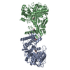
| ||||||||
|---|---|---|---|---|---|---|---|---|---|
| 1 |
| ||||||||
| Unit cell |
| ||||||||
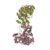
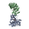
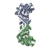
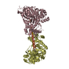
| Details | The biological assmbly is a dimer in the asymmetric unit. |
-Components
| #1: Protein | Mass: 55195.621 Da / Num. of mol.: 2 Source method: isolated from a genetically manipulated source Source: (gene. exp.) Cellulomonas sp. (bacteria) / Strain: NT3060 / Production host: #2: Chemical | ChemComp-MPD / (   Mass: 118.174 Da / Num. of mol.: 4 / Source method: obtained synthetically / Formula: C6H14O2 / Comment: precipitant*YM Mass: 118.174 Da / Num. of mol.: 4 / Source method: obtained synthetically / Formula: C6H14O2 / Comment: precipitant*YM#3: Water | ChemComp-HOH / |  Mass: 18.015 Da / Num. of mol.: 353 / Source method: isolated from a natural source / Formula: H2O Mass: 18.015 Da / Num. of mol.: 353 / Source method: isolated from a natural source / Formula: H2O |
|---|
-Experimental details
-Experiment
| Experiment | Method: X-RAY DIFFRACTION / Number of used crystals: 1 |
|---|
- Sample preparation
| Crystal | Density Matthews: 2.46 Å3/Da / Density % sol: 50.05 % |
|---|---|
| Crystal grow | Temperature: 277 K / Method: vapor diffusion / pH: 4.5 Details: 27-37% MPD, 0.1M citrate, pH 4.5, VAPOR DIFFUSION, temperature 277K |
-Data collection
| Diffraction | Mean temperature: 100 K |
|---|---|
| Diffraction source | Source: SYNCHROTRON / Site: SPring-8  / Beamline: BL41XU / Wavelength: 1.13 Å / Beamline: BL41XU / Wavelength: 1.13 Å |
| Detector | Type: MARRESEARCH / Detector: CCD / Date: Feb 5, 2003 |
| Radiation | Monochromator: double-crystal monochromator / Protocol: SINGLE WAVELENGTH / Monochromatic (M) / Laue (L): M / Scattering type: x-ray |
| Radiation wavelength | Wavelength: 1.13 Å / Relative weight: 1 |
| Reflection | Resolution: 2.3→55.9 Å / Num. all: 50338 / Num. obs: 49935 / % possible obs: 99.2 % / Observed criterion σ(F): 0 / Observed criterion σ(I): 0 / Redundancy: 9.1 % / Rmerge(I) obs: 0.104 / Net I/σ(I): 5.8 |
| Reflection shell | Resolution: 2.3→2.42 Å / % possible all: 99.2 |
- Processing
| Software |
| ||||||||||||||||||||||||||||||||||||||||||||||||||||||||||||||||||||||||||||||||||||||||||||||||||||||||||||||||||||||||||||||||||||||||||||||||||||||||||||||||||||||||||
|---|---|---|---|---|---|---|---|---|---|---|---|---|---|---|---|---|---|---|---|---|---|---|---|---|---|---|---|---|---|---|---|---|---|---|---|---|---|---|---|---|---|---|---|---|---|---|---|---|---|---|---|---|---|---|---|---|---|---|---|---|---|---|---|---|---|---|---|---|---|---|---|---|---|---|---|---|---|---|---|---|---|---|---|---|---|---|---|---|---|---|---|---|---|---|---|---|---|---|---|---|---|---|---|---|---|---|---|---|---|---|---|---|---|---|---|---|---|---|---|---|---|---|---|---|---|---|---|---|---|---|---|---|---|---|---|---|---|---|---|---|---|---|---|---|---|---|---|---|---|---|---|---|---|---|---|---|---|---|---|---|---|---|---|---|---|---|---|---|---|---|---|
| Refinement | Method to determine structure: SIROAS / Resolution: 2.3→20 Å / Cor.coef. Fo:Fc: 0.944 / Cor.coef. Fo:Fc free: 0.912 / SU B: 7.016 / SU ML: 0.174 / Cross valid method: THROUGHOUT / σ(F): 0 / ESU R: 0.338 / ESU R Free: 0.234 / Stereochemistry target values: MAXIMUM LIKELIHOOD
| ||||||||||||||||||||||||||||||||||||||||||||||||||||||||||||||||||||||||||||||||||||||||||||||||||||||||||||||||||||||||||||||||||||||||||||||||||||||||||||||||||||||||||
| Solvent computation | Ion probe radii: 0.8 Å / Shrinkage radii: 0.8 Å / VDW probe radii: 1.4 Å / Solvent model: BABINET MODEL WITH MASK | ||||||||||||||||||||||||||||||||||||||||||||||||||||||||||||||||||||||||||||||||||||||||||||||||||||||||||||||||||||||||||||||||||||||||||||||||||||||||||||||||||||||||||
| Displacement parameters | Biso mean: 30.327 Å2
| ||||||||||||||||||||||||||||||||||||||||||||||||||||||||||||||||||||||||||||||||||||||||||||||||||||||||||||||||||||||||||||||||||||||||||||||||||||||||||||||||||||||||||
| Refinement step | Cycle: LAST / Resolution: 2.3→20 Å
| ||||||||||||||||||||||||||||||||||||||||||||||||||||||||||||||||||||||||||||||||||||||||||||||||||||||||||||||||||||||||||||||||||||||||||||||||||||||||||||||||||||||||||
| Refine LS restraints |
| ||||||||||||||||||||||||||||||||||||||||||||||||||||||||||||||||||||||||||||||||||||||||||||||||||||||||||||||||||||||||||||||||||||||||||||||||||||||||||||||||||||||||||
| LS refinement shell | Resolution: 2.3→2.374 Å / Total num. of bins used: 16 /
|
