 Movie
Movie Controller


+ Open data

- Basic information
| Entry | Database: PDB / ID: 6iu3 | ||||||
|---|---|---|---|---|---|---|---|
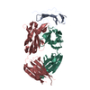
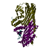
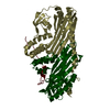
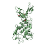
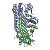
| Title | Crystal structure of iron transporter VIT1 with zinc ions | ||||||
 Components Components | VIT1 | ||||||
 Keywords Keywords | METAL TRANSPORT / membrane protein | ||||||
| Function / homology | (2R)-2,3-dihydroxypropyl (9Z)-octadec-9-enoate Function and homology information Function and homology information | ||||||
| Biological species |  Eucalyptus grandis (rose gum) Eucalyptus grandis (rose gum) | ||||||
| Method |  X-RAY DIFFRACTION / SYNCHROTRON / SAD / Resolution: 2.7 Å X-RAY DIFFRACTION / SYNCHROTRON / SAD / Resolution: 2.7 Å | ||||||
 Authors Authors | Kato, T. / Nishizawa, T. / Yamashita, K. / Taniguchi, R. / Kumazaki, K. / Ishitani, R. / Nureki, O. | ||||||
 Citation Citation | Journal: Nat Plants / Year: 2019 Title: Crystal structure of plant vacuolar iron transporter VIT1. Authors: Kato, T. / Kumazaki, K. / Wada, M. / Taniguchi, R. / Nakane, T. / Yamashita, K. / Hirata, K. / Ishitani, R. / Ito, K. / Nishizawa, T. / Nureki, O. | ||||||
| History |
|
- Structure visualization
| Structure viewer | Molecule: MolmilJmol/JSmol |
|---|
- Downloads & links
-Download
| PDBx/mmCIF format | 6iu3.cif.gz | 56.1 KB | Display | PDBx/mmCIF format |
|---|---|---|---|---|
| PDB format | pdb6iu3.ent.gz | 37.8 KB | Display | PDB format |
| PDBx/mmJSON format | 6iu3.json.gz | Tree view | PDBx/mmJSON format | |
| Others |  Other downloads Other downloads |
-Validation report
| Arichive directory | https://data.pdbj.org/pub/pdb/validation_reports/iu/6iu3ftp://data.pdbj.org/pub/pdb/validation_reports/iu/6iu3 | HTTPS FTP |
|---|
-Related structure data
| Related structure data | 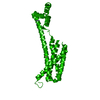 6iu4C 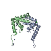 6iu5C  6iu6C  6iu8C  6iu9C C: citing same article ( |
|---|---|
| Similar structure data | |
| Experimental dataset #1 | Data reference: 10.5281/zenodo.2532136 / Data set type: diffraction image data |

-Links
 PDBj
PDBj


- Assembly
| Deposited unit | 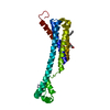
| ||||||||
|---|---|---|---|---|---|---|---|---|---|
| 1 | 
| ||||||||
| Unit cell |
|
-Components
| #1: Protein | Mass: 24960.010 Da / Num. of mol.: 1 Source method: isolated from a genetically manipulated source Source: (gene. exp.) Eucalyptus grandis (rose gum) / Plasmid: modified pYES2 / Production host:  |
|---|---|
| #2: Chemical | ChemComp-OLC / (  Mass: 356.540 Da / Num. of mol.: 1 / Source method: obtained synthetically / Formula: C21H40O4 Mass: 356.540 Da / Num. of mol.: 1 / Source method: obtained synthetically / Formula: C21H40O4 |
| #3: Chemical |   Mass: 65.409 Da / Num. of mol.: 3 / Source method: obtained synthetically / Formula: Zn Mass: 65.409 Da / Num. of mol.: 3 / Source method: obtained synthetically / Formula: Zn |
-Experimental details
-Experiment
| Experiment | Method: X-RAY DIFFRACTION / Number of used crystals: 1 |
|---|
- Sample preparation
| Crystal | Density Matthews: 3.13 Å3/Da / Density % sol: 60.66 % Description: The entry contains friedel pairs in I_plus/minus columns |
|---|---|
| Crystal grow | Temperature: 293 K / Method: lipidic cubic phase / pH: 7 Details: 24-26% PEG 500DME, 0.1 M HEPES pH7.0, 0.25-30 M ammonium sulfate, 0.0004 M zinc cloride, 0.2 M sodium fluoride |
-Data collection
| Diffraction | Mean temperature: 100 K / Serial crystal experiment: N | ||||||||||||||||||||||||||||||||||||||||||||||||||||||||||||||||||||||||||||||||||||||||
|---|---|---|---|---|---|---|---|---|---|---|---|---|---|---|---|---|---|---|---|---|---|---|---|---|---|---|---|---|---|---|---|---|---|---|---|---|---|---|---|---|---|---|---|---|---|---|---|---|---|---|---|---|---|---|---|---|---|---|---|---|---|---|---|---|---|---|---|---|---|---|---|---|---|---|---|---|---|---|---|---|---|---|---|---|---|---|---|---|---|
| Diffraction source | Source: SYNCHROTRON / Site: SPring-8  / Beamline: BL32XU / Wavelength: 1 Å / Beamline: BL32XU / Wavelength: 1 Å | ||||||||||||||||||||||||||||||||||||||||||||||||||||||||||||||||||||||||||||||||||||||||
| Detector | Type: DECTRIS EIGER X 9M / Detector: PIXEL / Date: Sep 26, 2016 | ||||||||||||||||||||||||||||||||||||||||||||||||||||||||||||||||||||||||||||||||||||||||
| Radiation | Monochromator: Si(111) / Protocol: SINGLE WAVELENGTH / Monochromatic (M) / Laue (L): M / Scattering type: x-ray | ||||||||||||||||||||||||||||||||||||||||||||||||||||||||||||||||||||||||||||||||||||||||
| Radiation wavelength | Wavelength: 1 Å / Relative weight: 1 | ||||||||||||||||||||||||||||||||||||||||||||||||||||||||||||||||||||||||||||||||||||||||
| Reflection | Resolution: 2.7001→48.375 Å / Num. obs: 16510 / % possible obs: 99.6154 % / Observed criterion σ(I): -3 / Redundancy: 12.782 % / Biso Wilson estimate: 74.36 Å2 / CC1/2: 0.9975 / Rmerge(I) obs: 0.1134 / Net I/σ(I): 14.5139 / Num. measured all: 115869 | ||||||||||||||||||||||||||||||||||||||||||||||||||||||||||||||||||||||||||||||||||||||||
| Reflection shell |
|
- Processing
| Software |
| |||||||||||||||||||||||||||||||||||||||||||||||||||||||||||||||||||||||||||||||||||||||||||
|---|---|---|---|---|---|---|---|---|---|---|---|---|---|---|---|---|---|---|---|---|---|---|---|---|---|---|---|---|---|---|---|---|---|---|---|---|---|---|---|---|---|---|---|---|---|---|---|---|---|---|---|---|---|---|---|---|---|---|---|---|---|---|---|---|---|---|---|---|---|---|---|---|---|---|---|---|---|---|---|---|---|---|---|---|---|---|---|---|---|---|---|---|
| Refinement | Method to determine structure: SAD / Resolution: 2.7→48.373 Å / SU ML: 0.51 / Cross valid method: THROUGHOUT / σ(F): 1.34 / Phase error: 37.13 / Stereochemistry target values: ML Details: The entry contains friedel pairs in I_plus/minus columns
| |||||||||||||||||||||||||||||||||||||||||||||||||||||||||||||||||||||||||||||||||||||||||||
| Solvent computation | Shrinkage radii: 0.9 Å / VDW probe radii: 1.11 Å / Solvent model: FLAT BULK SOLVENT MODEL | |||||||||||||||||||||||||||||||||||||||||||||||||||||||||||||||||||||||||||||||||||||||||||
| Displacement parameters | Biso max: 425.4 Å2 / Biso mean: 114.0346 Å2 / Biso min: 43.01 Å2 | |||||||||||||||||||||||||||||||||||||||||||||||||||||||||||||||||||||||||||||||||||||||||||
| Refinement step | Cycle: final / Resolution: 2.7→48.373 Å
| |||||||||||||||||||||||||||||||||||||||||||||||||||||||||||||||||||||||||||||||||||||||||||
| LS refinement shell | Refine-ID: X-RAY DIFFRACTION / Rfactor Rfree error: 0 / Total num. of bins used: 12
|
