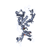
 Movie
Movie Controller


+ Open data

- Basic information
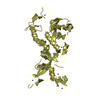
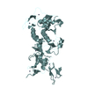
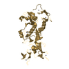
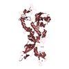
| Entry | Database: PDB / ID: 4k95 | ||||||
|---|---|---|---|---|---|---|---|
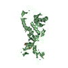
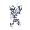
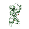
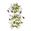
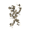
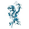
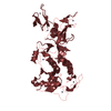
| Title | Crystal Structure of Parkin | ||||||
 Components Components | E3 ubiquitin-protein ligase parkin | ||||||
 Keywords Keywords | LIGASE / Ubiquitin-like domain / RING domains / zinc fingers / RBR ubiquitin ligase / zinc finger / UbcH7 / ubiquitin / mitochondria | ||||||
| Function / homology |  Function and homology information Function and homology informationJosephin domain DUBs / Regulation of necroptotic cell death / Aggrephagy / PINK1-PRKN Mediated Mitophagy / positive regulation of neurotransmitter uptake / negative regulation of endoplasmic reticulum stress-induced neuron intrinsic apoptotic signaling pathway / Antigen processing: Ubiquitination & Proteasome degradation / negative regulation of spontaneous neurotransmitter secretion / mitochondrion-derived vesicle / negative regulation of intralumenal vesicle formation ...Josephin domain DUBs / Regulation of necroptotic cell death / Aggrephagy / PINK1-PRKN Mediated Mitophagy / positive regulation of neurotransmitter uptake / negative regulation of endoplasmic reticulum stress-induced neuron intrinsic apoptotic signaling pathway / Antigen processing: Ubiquitination & Proteasome degradation / negative regulation of spontaneous neurotransmitter secretion / mitochondrion-derived vesicle / negative regulation of intralumenal vesicle formation / regulation protein catabolic process at presynapse / cellular response to L-glutamine / negative regulation of exosomal secretion / mitochondrion to lysosome vesicle-mediated transport / type 2 mitophagy / response to curcumin / free ubiquitin chain polymerization / negative regulation of mitochondrial fusion / cellular response to hydrogen sulfide / positive regulation of protein linear polyubiquitination / RBR-type E3 ubiquitin transferase / host-mediated suppression of viral genome replication / Parkin-FBXW7-Cul1 ubiquitin ligase complex / positive regulation of mitochondrial fusion / negative regulation of actin filament bundle assembly / mitochondrial fragmentation involved in apoptotic process / F-box domain binding / positive regulation of mitophagy / regulation of cellular response to oxidative stress / positive regulation of dendrite extension / regulation of neurotransmitter secretion / regulation of dopamine metabolic process / negative regulation of excitatory postsynaptic potential / norepinephrine metabolic process / autophagy of mitochondrion / positive regulation of type 2 mitophagy / dopaminergic synapse / mitochondrion localization / protein localization to mitochondrion / mitochondrial fission / negative regulation of intrinsic apoptotic signaling pathway by p53 class mediator / cellular response to toxic substance / positive regulation of tumor necrosis factor-mediated signaling pathway / negative regulation of oxidative stress-induced neuron intrinsic apoptotic signaling pathway / positive regulation of protein localization to membrane / regulation of mitochondrion organization / aggresome assembly / protein K11-linked ubiquitination / cellular response to L-glutamate / synaptic transmission, dopaminergic / positive regulation of proteasomal protein catabolic process / protein K6-linked ubiquitination / ubiquitin conjugating enzyme binding / aggresome / negative regulation of JNK cascade / negative regulation of synaptic transmission, glutamatergic / positive regulation of mitochondrial membrane potential / regulation of reactive oxygen species metabolic process / positive regulation of mitochondrial fission / response to muscle activity / negative regulation of release of cytochrome c from mitochondria / response to corticosterone / ubiquitin-specific protease binding / intracellular vesicle / startle response / dopamine metabolic process / dopamine uptake involved in synaptic transmission / negative regulation of reactive oxygen species metabolic process / positive regulation of ATP biosynthetic process / regulation of protein ubiquitination / response to unfolded protein / cullin family protein binding / regulation of synaptic vesicle endocytosis / negative regulation of mitochondrial fission / protein K63-linked ubiquitination / protein monoubiquitination / negative regulation of endoplasmic reticulum stress-induced intrinsic apoptotic signaling pathway / ubiquitin ligase complex / regulation of postsynaptic membrane neurotransmitter receptor levels / protein autoubiquitination / mitophagy / phospholipase binding / negative regulation of reactive oxygen species biosynthetic process / cellular response to manganese ion / heat shock protein binding / protein K48-linked ubiquitination / proteasomal protein catabolic process / Hsp70 protein binding / response to endoplasmic reticulum stress / positive regulation of insulin secretion involved in cellular response to glucose stimulus / protein catabolic process / regulation of autophagy / learning / regulation of mitochondrial membrane potential / adult locomotory behavior / PDZ domain binding / ubiquitin binding / mitochondrion organization / regulation of protein stability / synaptic transmission, glutamatergic Similarity search - Function | ||||||
| Biological species |  | ||||||
| Method |  X-RAY DIFFRACTION / SYNCHROTRON / MOLECULAR REPLACEMENT / Resolution: 6.499 Å X-RAY DIFFRACTION / SYNCHROTRON / MOLECULAR REPLACEMENT / Resolution: 6.499 Å | ||||||
 Authors Authors | Seirafi, M. / Menade, M. / Sauve, V. / Kozlov, G. / Trempe, J.-F. / Nagar, B. / Gehring, K. | ||||||
 Citation Citation | Journal: Science / Year: 2013 Title: Structure of parkin reveals mechanisms for ubiquitin ligase activation. Authors: Trempe, J.F. / Sauve, V. / Grenier, K. / Seirafi, M. / Tang, M.Y. / Menade, M. / Al-Abdul-Wahid, S. / Krett, J. / Wong, K. / Kozlov, G. / Nagar, B. / Fon, E.A. / Gehring, K. | ||||||
| History |
|
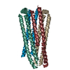
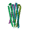
- Structure visualization
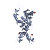
| Structure viewer | Molecule: MolmilJmol/JSmol |
|---|
- Downloads & links
-Download
| PDBx/mmCIF format | 4k95.cif.gz | 778.1 KB | Display | PDBx/mmCIF format |
|---|---|---|---|---|
| PDB format | pdb4k95.ent.gz | 621.4 KB | Display | PDB format |
| PDBx/mmJSON format | 4k95.json.gz | Tree view | PDBx/mmJSON format | |
| Others |  Other downloads Other downloads |
-Validation report
| Arichive directory | https://data.pdbj.org/pub/pdb/validation_reports/k9/4k95ftp://data.pdbj.org/pub/pdb/validation_reports/k9/4k95 | HTTPS FTP |
|---|
-Related structure data
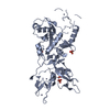
| Related structure data | 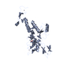 4k7dSC 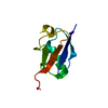 2zeqS S: Starting model for refinement C: citing same article ( |
|---|---|
| Similar structure data |

-Links
 PDBj
PDBj

- Assembly


-Components
| #1: Protein | Mass: 52155.113 Da / Num. of mol.: 12 Source method: isolated from a genetically manipulated source Details: Codon-optimized for E.coli expression / Source: (gene. exp.)  References: UniProt: Q9JK66, Ligases; Forming carbon-nitrogen bonds; Acid-amino-acid ligases (peptide synthases) #2: Chemical | ChemComp-ZN /   Mass: 65.409 Da / Num. of mol.: 96 / Source method: obtained synthetically / Formula: Zn Mass: 65.409 Da / Num. of mol.: 96 / Source method: obtained synthetically / Formula: Zn |
|---|
-Experimental details
-Experiment
| Experiment | Method: X-RAY DIFFRACTION / Number of used crystals: 1 |
|---|
- Sample preparation
| Crystal | Density Matthews: 2.91 Å3/Da / Density % sol: 57.74 % |
|---|---|
| Crystal grow | Temperature: 277 K / Method: vapor diffusion, hanging drop / pH: 7 Details: 22.5% PEG3350, 0.2 M (NH4)2SO4, 0.1 M HEPES pH 7.0, 10% (v/v) glycerol, 3% sorbitol and 25 mM 2-mercaptoethanol, vapor diffusion, hanging drop, temperature 277K |
-Data collection
| Diffraction | Mean temperature: 100 K | |||||||||||||||||||||||||||||||||||||||||||||||||||||||||||||||||||||||||||||
|---|---|---|---|---|---|---|---|---|---|---|---|---|---|---|---|---|---|---|---|---|---|---|---|---|---|---|---|---|---|---|---|---|---|---|---|---|---|---|---|---|---|---|---|---|---|---|---|---|---|---|---|---|---|---|---|---|---|---|---|---|---|---|---|---|---|---|---|---|---|---|---|---|---|---|---|---|---|---|
| Diffraction source | Source: SYNCHROTRON / Site: CHESS  / Beamline: F2 / Wavelength: 0.9183 Å / Beamline: F2 / Wavelength: 0.9183 Å | |||||||||||||||||||||||||||||||||||||||||||||||||||||||||||||||||||||||||||||
| Detector | Type: ADSC QUANTUM 210 / Detector: CCD / Date: Jun 11, 2012 / Details: mirrors | |||||||||||||||||||||||||||||||||||||||||||||||||||||||||||||||||||||||||||||
| Radiation | Monochromator: Si(111) / Protocol: SINGLE WAVELENGTH / Monochromatic (M) / Laue (L): M / Scattering type: x-ray | |||||||||||||||||||||||||||||||||||||||||||||||||||||||||||||||||||||||||||||
| Radiation wavelength | Wavelength: 0.9183 Å / Relative weight: 1 | |||||||||||||||||||||||||||||||||||||||||||||||||||||||||||||||||||||||||||||
| Reflection | Resolution: 6.5→50 Å / Num. all: 14952 / Num. obs: 14922 / % possible obs: 99.8 % / Observed criterion σ(F): 1 / Observed criterion σ(I): 1 / Redundancy: 7.2 % / Biso Wilson estimate: 227 Å2 / Rmerge(I) obs: 0.172 / Χ2: 1.574 / Net I/σ(I): 15.1 | |||||||||||||||||||||||||||||||||||||||||||||||||||||||||||||||||||||||||||||
| Reflection shell | Diffraction-ID: 1
|
- Processing
| Software |
| ||||||||||||||||||||||||||||||||||||||||||||||||
|---|---|---|---|---|---|---|---|---|---|---|---|---|---|---|---|---|---|---|---|---|---|---|---|---|---|---|---|---|---|---|---|---|---|---|---|---|---|---|---|---|---|---|---|---|---|---|---|---|---|
| Refinement | Method to determine structure: MOLECULAR REPLACEMENT Starting model: PDB entries: 4K7D (rat parkin 141-465) and 2ZEQ (mouse parkin 1-72) Resolution: 6.499→49.334 Å / Occupancy max: 1 / Occupancy min: 0.5 / FOM work R set: 0.7121 / SU ML: 1.11 / σ(F): 1.34 / Phase error: 34.11 / Stereochemistry target values: ML
| ||||||||||||||||||||||||||||||||||||||||||||||||
| Solvent computation | Shrinkage radii: 0.9 Å / VDW probe radii: 1.11 Å / Solvent model: FLAT BULK SOLVENT MODEL | ||||||||||||||||||||||||||||||||||||||||||||||||
| Displacement parameters | Biso max: 213.59 Å2 / Biso mean: 88.6785 Å2 / Biso min: 28.1 Å2 | ||||||||||||||||||||||||||||||||||||||||||||||||
| Refinement step | Cycle: LAST / Resolution: 6.499→49.334 Å
| ||||||||||||||||||||||||||||||||||||||||||||||||
| Refine LS restraints |
| ||||||||||||||||||||||||||||||||||||||||||||||||
| LS refinement shell | Refine-ID: X-RAY DIFFRACTION / Total num. of bins used: 5
|
