positive regulation of retrograde transport, endosome to Golgi / regulation of lipid transport / positive regulation of neurotransmitter uptake / negative regulation of endoplasmic reticulum stress-induced neuron intrinsic apoptotic signaling pathway / negative regulation of spontaneous neurotransmitter secretion / negative regulation of intralumenal vesicle formation / regulation protein catabolic process at presynapse / cellular response to L-glutamine / : / negative regulation of exosomal secretion ...positive regulation of retrograde transport, endosome to Golgi / regulation of lipid transport / positive regulation of neurotransmitter uptake / negative regulation of endoplasmic reticulum stress-induced neuron intrinsic apoptotic signaling pathway / negative regulation of spontaneous neurotransmitter secretion / negative regulation of intralumenal vesicle formation / regulation protein catabolic process at presynapse / cellular response to L-glutamine / : / negative regulation of exosomal secretion / mitochondrion to lysosome vesicle-mediated transport / type 2 mitophagy / negative regulation of glucokinase activity / response to curcumin / protein K29-linked ubiquitination / free ubiquitin chain polymerization / negative regulation of mitochondrial fusion / cellular response to hydrogen sulfide / positive regulation of protein linear polyubiquitination / RBR-type E3 ubiquitin transferase / regulation of synaptic vesicle transport / host-mediated suppression of viral genome replication / Parkin-FBXW7-Cul1 ubiquitin ligase complex / positive regulation of mitochondrial fusion / negative regulation of actin filament bundle assembly / mitochondrial fragmentation involved in apoptotic process / regulation of necroptotic process / F-box domain binding / positive regulation of mitophagy / regulation of cellular response to oxidative stress / positive regulation of dendrite extension / regulation of dopamine metabolic process / negative regulation of excitatory postsynaptic potential / autophagy of mitochondrion / positive regulation of type 2 mitophagy / dopaminergic synapse / mitochondrion localization / protein localization to mitochondrion / cellular response to dopamine / mitochondrial fission / negative regulation of intrinsic apoptotic signaling pathway by p53 class mediator / cellular response to toxic substance / positive regulation of tumor necrosis factor-mediated signaling pathway / negative regulation of oxidative stress-induced neuron intrinsic apoptotic signaling pathway / positive regulation of protein localization to membrane / regulation of mitochondrion organization / aggresome assembly / protein K11-linked ubiquitination / protein K6-linked ubiquitination / cellular response to L-glutamate / aggresome / positive regulation of proteasomal protein catabolic process / negative regulation of JNK cascade / ubiquitin conjugating enzyme binding / regulation of canonical Wnt signaling pathway / regulation of reactive oxygen species metabolic process / positive regulation of mitochondrial membrane potential / negative regulation of synaptic transmission, glutamatergic / protein K27-linked ubiquitination / positive regulation of mitochondrial fission / response to muscle activity / negative regulation of release of cytochrome c from mitochondria / response to corticosterone / Lewy body / ubiquitin-specific protease binding / dopamine metabolic process / regulation of dopamine secretion / regulation of glucose metabolic process / negative regulation of reactive oxygen species metabolic process / positive regulation of ATP biosynthetic process / regulation of protein ubiquitination / cellular response to unfolded protein / cullin family protein binding / regulation of synaptic vesicle endocytosis / negative regulation of mitochondrial fission / protein monoubiquitination / negative regulation of insulin secretion / protein deubiquitination / protein K63-linked ubiquitination / negative regulation of endoplasmic reticulum stress-induced intrinsic apoptotic signaling pathway / ubiquitin ligase complex / regulation of postsynaptic membrane neurotransmitter receptor levels / protein autoubiquitination / mitophagy / negative regulation of reactive oxygen species biosynthetic process / phospholipase binding / adult locomotory behavior / cellular response to manganese ion / heat shock protein binding / ERAD pathway / protein K48-linked ubiquitination / Hsp70 protein binding / response to endoplasmic reticulum stress / positive regulation of insulin secretion involved in cellular response to glucose stimulus / regulation of autophagy / Josephin domain DUBs / central nervous system development / PINK1-PRKN Mediated Mitophagy / macroautophagy / proteasomal protein catabolic process Similarity search - Function
Resolution: 2.85→47.98 Å / Cor.coef. Fo:Fc: 0.929 / Cor.coef. Fo:Fc free: 0.913 / SU B: 24.598 / SU ML: 0.407 / Cross valid method: THROUGHOUT / ESU R Free: 0.422 / Details: HYDROGENS HAVE BEEN ADDED IN THE RIDING POSITIONS
Rfactor
Num. reflection
% reflection
Selection details
Rfree
0.25486
1095
5.2 %
RANDOM
Rwork
0.22284
-
-
-
obs
0.22449
20094
95.04 %
-
Solvent computation
Ion probe radii: 0.8 Å / Shrinkage radii: 0.8 Å / VDW probe radii: 1.2 Å
 Movie
Movie Controller


 Open data
 Basic information
 Components
 Keywords
 Function and homology information
 Homo sapiens (human)
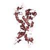
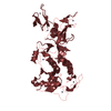
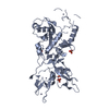
 X-RAY DIFFRACTION /
  Authors
 United Kingdom, 1items
  Citation
 Structure visualization
 Downloads & links
 Other downloads
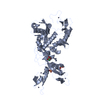


 PDBj
PDBj

 Assembly
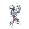
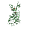


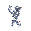
 Mass: 65.409 Da / Num. of mol.: 16 / Source method: obtained synthetically / Formula: Zn
 Mass: 96.063 Da / Num. of mol.: 2 / Source method: obtained synthetically / Formula: SO4
 Mass: 92.094 Da / Num. of mol.: 4 / Source method: obtained synthetically / Formula: C3H8O3
 Mass: 35.453 Da / Num. of mol.: 2 / Source method: obtained synthetically / Formula: Cl
 Sample preparation
 / Beamline: MASSIF-1 / Wavelength: 0.966 Å
 Processing