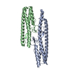
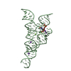
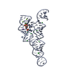
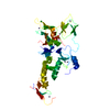
[English] 日本語
Yorodumi
- PDB-4k7d: Crystal Structure of Parkin C-terminal RING domains -

+
Open data


ID or keywords:

Loading...

-
Basic information

Entry
Database: PDB / ID: 4k7d
TitleCrystal Structure of Parkin C-terminal RING domains
ComponentsE3 ubiquitin-protein ligase parkin
KeywordsLIGASE / RING domains / zinc fingers / RBR ubiquitin ligase / E3 ubiquitin protein ligase / Ubiquitin / UbcH7
Function / homology
Function and homology information


Josephin domain DUBs / Regulation of necroptotic cell death / Aggrephagy / PINK1-PRKN Mediated Mitophagy / cellular response to L-glutamine / positive regulation of neurotransmitter uptake / negative regulation of endoplasmic reticulum stress-induced neuron intrinsic apoptotic signaling pathway / Antigen processing: Ubiquitination & Proteasome degradation / mitochondrion-derived vesicle / negative regulation of spontaneous neurotransmitter secretion ...Josephin domain DUBs / Regulation of necroptotic cell death / Aggrephagy / PINK1-PRKN Mediated Mitophagy / cellular response to L-glutamine / positive regulation of neurotransmitter uptake / negative regulation of endoplasmic reticulum stress-induced neuron intrinsic apoptotic signaling pathway / Antigen processing: Ubiquitination & Proteasome degradation / mitochondrion-derived vesicle / negative regulation of spontaneous neurotransmitter secretion / negative regulation of intralumenal vesicle formation / regulation protein catabolic process at presynapse / response to curcumin / negative regulation of exosomal secretion / negative regulation of glucokinase activity / mitochondrion to lysosome vesicle-mediated transport / cellular response to hydrogen sulfide / type 2 mitophagy / negative regulation of actin filament bundle assembly / negative regulation of mitochondrial fusion / Parkin-FBXW7-Cul1 ubiquitin ligase complex / free ubiquitin chain polymerization / positive regulation of protein linear polyubiquitination / negative regulation by host of viral genome replication / positive regulation of mitophagy / RBR-type E3 ubiquitin transferase / mitochondrial fragmentation involved in apoptotic process / F-box domain binding / positive regulation of mitochondrial fusion / mitochondrion localization / regulation of cellular response to oxidative stress / negative regulation of excitatory postsynaptic potential / regulation of dopamine metabolic process / regulation of neurotransmitter secretion / negative regulation of intrinsic apoptotic signaling pathway by p53 class mediator / dopaminergic synapse / positive regulation of dendrite extension / protein K6-linked ubiquitination / positive regulation of protein localization to membrane / cellular response to L-glutamate / autophagy of mitochondrion / norepinephrine metabolic process / cellular response to toxic substance / positive regulation of type 2 mitophagy / protein localization to mitochondrion / positive regulation of proteasomal protein catabolic process / synaptic transmission, dopaminergic / negative regulation of JNK cascade / mitochondrial fission / positive regulation of tumor necrosis factor-mediated signaling pathway / negative regulation of synaptic transmission, glutamatergic / negative regulation of oxidative stress-induced neuron intrinsic apoptotic signaling pathway / aggresome assembly / protein K11-linked ubiquitination / ubiquitin conjugating enzyme binding / regulation of mitochondrion organization / positive regulation of mitochondrial membrane potential / aggresome / regulation of reactive oxygen species metabolic process / response to corticosterone / positive regulation of mitochondrial fission / positive regulation of ATP biosynthetic process / response to muscle activity / dopamine uptake involved in synaptic transmission / intracellular vesicle / ubiquitin-specific protease binding / negative regulation of release of cytochrome c from mitochondria / startle response / dopamine metabolic process / phospholipase binding / regulation of synaptic vesicle endocytosis / cullin family protein binding / negative regulation of reactive oxygen species metabolic process / positive regulation of insulin secretion involved in cellular response to glucose stimulus / protein K63-linked ubiquitination / negative regulation of mitochondrial fission / protein monoubiquitination / regulation of protein ubiquitination / cellular response to manganese ion / ubiquitin ligase complex / response to unfolded protein / regulation of postsynaptic membrane neurotransmitter receptor levels / negative regulation of insulin secretion / proteasomal protein catabolic process / protein K48-linked ubiquitination / negative regulation of endoplasmic reticulum stress-induced intrinsic apoptotic signaling pathway / protein autoubiquitination / mitophagy / negative regulation of reactive oxygen species biosynthetic process / heat shock protein binding / Hsp70 protein binding / tubulin binding / response to endoplasmic reticulum stress / regulation of mitochondrial membrane potential / adult locomotory behavior / ubiquitin binding / learning / synaptic transmission, glutamatergic / PDZ domain binding / mitochondrion organization
Similarity search - Function
: / E3 ubiquitin-protein ligase parkin / RING/Ubox-like zinc-binding domain / Parkin, RING/Ubox like zinc-binding domain / : / : / : / RING/Ubox like zinc-binding domain / RING/Ubox like zinc-binding domain / IBR domain ...: / E3 ubiquitin-protein ligase parkin / RING/Ubox-like zinc-binding domain / Parkin, RING/Ubox like zinc-binding domain / : / : / : / RING/Ubox like zinc-binding domain / RING/Ubox like zinc-binding domain / IBR domain / IBR domain, a half RING-finger domain / : / IBR domain / In Between Ring fingers / TRIAD supradomain / TRIAD supradomain profile. / Ubiquitin family / Ubiquitin homologues / Ubiquitin domain profile. / Ubiquitin-like domain / Ubiquitin-like domain superfamily
Similarity search - Domain/homology
MALONATE ION / E3 ubiquitin-protein ligase parkin
Similarity search - Component
Biological speciesRattus norvegicus (Norway rat)
MethodX-RAY DIFFRACTION / SYNCHROTRON / SAD / Resolution: 2.8 Å
AuthorsSauve, V. / Trempe, J.-F. / Menade, M. / Gehring, K.
CitationJournal: Science / Year: 2013
Title: Structure of parkin reveals mechanisms for ubiquitin ligase activation.
Authors: Trempe, J.F. / Sauve, V. / Grenier, K. / Seirafi, M. / Tang, M.Y. / Menade, M. / Al-Abdul-Wahid, S. / Krett, J. / Wong, K. / Kozlov, G. / Nagar, B. / Fon, E.A. / Gehring, K.
History
DepositionApr 17, 2013Deposition site: RCSB / Processing site: RCSB
Revision 1.0May 15, 2013Provider: repository / Type: Initial release
Revision 1.1May 22, 2013Group: Database references
Revision 1.2Jul 10, 2013Group: Database references
Revision 1.3Feb 28, 2024Group: Data collection / Database references / Derived calculations
Category: chem_comp_atom / chem_comp_bond ...chem_comp_atom / chem_comp_bond / database_2 / pdbx_struct_conn_angle / struct_conn / struct_ref_seq_dif / struct_site
Item: _database_2.pdbx_DOI / _database_2.pdbx_database_accession ..._database_2.pdbx_DOI / _database_2.pdbx_database_accession / _pdbx_struct_conn_angle.ptnr1_auth_asym_id / _pdbx_struct_conn_angle.ptnr1_auth_comp_id / _pdbx_struct_conn_angle.ptnr1_auth_seq_id / _pdbx_struct_conn_angle.ptnr1_label_asym_id / _pdbx_struct_conn_angle.ptnr1_label_atom_id / _pdbx_struct_conn_angle.ptnr1_label_comp_id / _pdbx_struct_conn_angle.ptnr1_label_seq_id / _pdbx_struct_conn_angle.ptnr2_auth_asym_id / _pdbx_struct_conn_angle.ptnr2_auth_seq_id / _pdbx_struct_conn_angle.ptnr2_label_asym_id / _pdbx_struct_conn_angle.ptnr3_auth_asym_id / _pdbx_struct_conn_angle.ptnr3_auth_comp_id / _pdbx_struct_conn_angle.ptnr3_auth_seq_id / _pdbx_struct_conn_angle.ptnr3_label_asym_id / _pdbx_struct_conn_angle.ptnr3_label_atom_id / _pdbx_struct_conn_angle.ptnr3_label_comp_id / _pdbx_struct_conn_angle.ptnr3_label_seq_id / _pdbx_struct_conn_angle.value / _struct_conn.pdbx_dist_value / _struct_conn.ptnr1_auth_asym_id / _struct_conn.ptnr1_auth_comp_id / _struct_conn.ptnr1_auth_seq_id / _struct_conn.ptnr1_label_asym_id / _struct_conn.ptnr1_label_atom_id / _struct_conn.ptnr1_label_comp_id / _struct_conn.ptnr1_label_seq_id / _struct_conn.ptnr2_auth_asym_id / _struct_conn.ptnr2_auth_seq_id / _struct_conn.ptnr2_label_asym_id / _struct_ref_seq_dif.details / _struct_site.pdbx_auth_asym_id / _struct_site.pdbx_auth_comp_id / _struct_site.pdbx_auth_seq_id

-
Structure visualization

Structure viewerMolecule:
MolmilJmol/JSmol

Downloads & links

-
Assembly

Deposited unit
A: E3 ubiquitin-protein ligase parkin
B: E3 ubiquitin-protein ligase parkin
C: E3 ubiquitin-protein ligase parkin
hetero molecules


Theoretical massNumber of molelcules
Total (without water)112,40531
Polymers110,4933
Non-polymers1,91128
Water81145
1
A: E3 ubiquitin-protein ligase parkin
hetero molecules


Theoretical massNumber of molelcules
Total (without water)37,45610
Polymers36,8311
Non-polymers6259
Water181
TypeNameSymmetry operationNumber
identity operation1_555x,y,z1
MethodPISA
2
B: E3 ubiquitin-protein ligase parkin
hetero molecules


Theoretical massNumber of molelcules
Total (without water)37,45610
Polymers36,8311
Non-polymers6259
Water181
TypeNameSymmetry operationNumber
identity operation1_555x,y,z1
MethodPISA
3
C: E3 ubiquitin-protein ligase parkin
hetero molecules


Theoretical massNumber of molelcules
Total (without water)37,49211
Polymers36,8311
Non-polymers66110
Water181
TypeNameSymmetry operationNumber
identity operation1_555x,y,z1
MethodPISA
Unit cell
Length a, b, c (Å)84.700, 106.625, 154.216
Angle α, β, γ (deg.)90.000, 90.000, 90.000
Int Tables number19
Space group name H-MP212121

-
Components

#1: Protein E3 ubiquitin-protein ligase parkin


Mass: 36831.070 Da / Num. of mol.: 3 / Fragment: C-terminal RING domains (141-465)
Source method: isolated from a genetically manipulated source
Details: Codon-optimized for E.coli expression / Source: (gene. exp.) Rattus norvegicus (Norway rat) / Gene: Park2, Prkn / Plasmid: pGEX-6P-1 / Production host: Escherichia coli (E. coli) / Strain (production host): BL21(DE3)
References: UniProt: Q9JK66, Ligases; Forming carbon-nitrogen bonds; Acid-amino-acid ligases (peptide synthases)
#2: Chemical...
ChemComp-ZN / ZINC ION


Mass: 65.409 Da / Num. of mol.: 24 / Source method: obtained synthetically / Formula: Zn
#3: Chemical ChemComp-MLI / MALONATE ION


Mass: 102.046 Da / Num. of mol.: 3 / Source method: obtained synthetically / Formula: C3H2O4
#4: Chemical ChemComp-CL / CHLORIDE ION


Mass: 35.453 Da / Num. of mol.: 1 / Source method: obtained synthetically / Formula: Cl
#5: Water ChemComp-HOH / water


Mass: 18.015 Da / Num. of mol.: 45 / Source method: isolated from a natural source / Formula: H2O

-
Experimental details

-
Experiment

ExperimentMethod: X-RAY DIFFRACTION / Number of used crystals: 1

-
Sample preparation

CrystalDensity Matthews: 3.15 Å3/Da / Density % sol: 60.97 %
Crystal growTemperature: 277 K / Method: vapor diffusion, hanging drop / pH: 7
Details: 0.9 M sodium malonate pH 7.0, 0.1 M HEPES pH 7.0, 5% (v/v) glycerol, 56 mM 2-mercaptoethanol, VAPOR DIFFUSION, HANGING DROP, temperature 277K

-
Data collection

DiffractionMean temperature: 100 K
Diffraction sourceSource: SYNCHROTRON / Site: CLSI / Beamline: 08ID-1 / Wavelength: 1.2824 Å
DetectorType: MARMOSAIC 300 mm CCD / Detector: CCD / Date: May 16, 2012 / Details: Vertical focusing mirrors
RadiationMonochromator: ACCEL/BRUKER DCM, Si(111) / Protocol: SINGLE WAVELENGTH / Monochromatic (M) / Laue (L): M / Scattering type: x-ray
Radiation wavelengthWavelength: 1.2824 Å / Relative weight: 1
ReflectionResolution: 2.8→53.33 Å / Num. all: 35155 / Num. obs: 35121 / % possible obs: 99.9 % / Observed criterion σ(F): 1 / Observed criterion σ(I): 1 / Redundancy: 9.7 % / Rmerge(I) obs: 0.079 / Net I/σ(I): 19.9
Reflection shell

Diffraction-ID: 1

Resolution (Å)Redundancy (%)Rmerge(I) obsMean I/σ(I) obsNum. measured allNum. unique allRsym value% possible all
2.8-2.959.70.4641.64874850440.464100
2.95-3.139.70.2792.64685248060.279100
3.13-3.359.80.154.94379644880.15100
3.35-3.619.80.0947.44111642160.094100
3.61-3.969.80.085.73805639010.08100
3.96-4.439.70.05411.53436035360.054100
4.43-5.119.70.05610.73045331440.056100
5.11-6.269.60.0728.12570526790.07299.8
6.26-8.859.50.04214.11985421030.04299.6
8.85-53.3138.90.03616.11081712380.03698.3

-
Phasing

PhasingMethod: SAD

-
Processing

Software
NameVersionClassificationNB
MOSFLMdata reduction
SCALA3.3.20data scaling
SOLVEphasing
RESOLVEphasing
PHENIX1.8.2_1309refinement
PDB_EXTRACT3.11data extraction
MxDCdata collection
PHENIX1.8.2_1309phasing
RefinementMethod to determine structure: SAD / Resolution: 2.8→53.31 Å / Occupancy max: 1 / Occupancy min: 0.5 / FOM work R set: 0.8301 / SU ML: 0.32 / σ(F): 1.36 / Phase error: 23.44 / Stereochemistry target values: ML
RfactorNum. reflection% reflectionSelection details
Rfree0.233 1782 5.08 %RANDOM
Rwork0.1844 ---
all0.187 35131 --
obs0.1869 35065 99.81 %-
Solvent computationShrinkage radii: 0.9 Å / VDW probe radii: 1.11 Å / Solvent model: FLAT BULK SOLVENT MODEL
Displacement parametersBiso max: 126.57 Å2 / Biso mean: 53.4322 Å2 / Biso min: 22.23 Å2
Refine analyzeLuzzati coordinate error obs: 0.337 Å
Refinement stepCycle: LAST / Resolution: 2.8→53.31 Å
ProteinNucleic acidLigandSolventTotal
Num. atoms7195 0 46 45 7286
Refine LS restraints
Refine-IDTypeDev idealNumber
X-RAY DIFFRACTIONf_bond_d0.017550
X-RAY DIFFRACTIONf_angle_d0.91410238
X-RAY DIFFRACTIONf_chiral_restr0.0661031
X-RAY DIFFRACTIONf_plane_restr0.0041360
X-RAY DIFFRACTIONf_dihedral_angle_d14.6772777
LS refinement shell

Refine-ID: X-RAY DIFFRACTION / Total num. of bins used: 13

Resolution (Å)Rfactor RfreeNum. reflection RfreeRfactor RworkNum. reflection RworkNum. reflection allNum. reflection obs% reflection obs (%)
2.7994-2.87510.31811330.2863248826212488100
2.8751-2.95970.29011330.242255426872554100
2.9597-3.05520.30291350.2276251226472512100
3.0552-3.16440.28341250.2256254826732548100
3.1644-3.2910.30351410.2193254226832542100
3.291-3.44080.29591340.2129252526592525100
3.4408-3.62220.23111450.1875252426692524100
3.6222-3.84910.22991310.1792257427052574100
3.8491-4.14610.2081350.1636254526802545100
4.1461-4.56320.17311410.1505257827192578100
4.5632-5.2230.19651460.1517258427302584100
5.223-6.57850.22721430.1721259927422599100
6.5785-53.32210.22091400.181727102850271099

+
About Yorodumi

-
News

-
Feb 9, 2022. New format data for meta-information of EMDB entries

New format data for meta-information of EMDB entries

  • Version 3 of the EMDB header file is now the official format.
  • The previous official version 1.9 will be removed from the archive.

Related info.:EMDB header

External links:wwPDB to switch to version 3 of the EMDB data model

-
Aug 12, 2020. Covid-19 info

Covid-19 info

URL: https://pdbj.org/emnavi/covid19.php

New page: Covid-19 featured information page in EM Navigator.

Related info.:Covid-19 info / Mar 5, 2020. Novel coronavirus structure data

+
Mar 5, 2020. Novel coronavirus structure data

Novel coronavirus structure data

Related info.:Yorodumi Speices / Aug 12, 2020. Covid-19 info

External links:COVID-19 featured content - PDBj / Molecule of the Month (242):Coronavirus Proteases

+
Jan 31, 2019. EMDB accession codes are about to change! (news from PDBe EMDB page)

EMDB accession codes are about to change! (news from PDBe EMDB page)

  • The allocation of 4 digits for EMDB accession codes will soon come to an end. Whilst these codes will remain in use, new EMDB accession codes will include an additional digit and will expand incrementally as the available range of codes is exhausted. The current 4-digit format prefixed with “EMD-” (i.e. EMD-XXXX) will advance to a 5-digit format (i.e. EMD-XXXXX), and so on. It is currently estimated that the 4-digit codes will be depleted around Spring 2019, at which point the 5-digit format will come into force.
  • The EM Navigator/Yorodumi systems omit the EMD- prefix.

Related info.:Q: What is EMD? / ID/Accession-code notation in Yorodumi/EM Navigator

External links:EMDB Accession Codes are Changing Soon! / Contact to PDBj

+
Jul 12, 2017. Major update of PDB

Major update of PDB

  • wwPDB released updated PDB data conforming to the new PDBx/mmCIF dictionary.
  • This is a major update changing the version number from 4 to 5, and with Remediation, in which all the entries are updated.
  • In this update, many items about electron microscopy experimental information are reorganized (e.g. em_software).
  • Now, EM Navigator and Yorodumi are based on the updated data.

External links:wwPDB Remediation / Enriched Model Files Conforming to OneDep Data Standards Now Available in the PDB FTP Archive

-
Yorodumi

Thousand views of thousand structures

  • Yorodumi is a browser for structure data from EMDB, PDB, SASBDB, etc.
  • This page is also the successor to EM Navigator detail page, and also detail information page/front-end page for Omokage search.
  • The word "yorodu" (or yorozu) is an old Japanese word meaning "ten thousand". "mi" (miru) is to see.

Related info.:EMDB / PDB / SASBDB / Comparison of 3 databanks / Yorodumi Search / Aug 31, 2016. New EM Navigator & Yorodumi / Yorodumi Papers / Jmol/JSmol / Function and homology information / Changes in new EM Navigator and Yorodumi

Read more