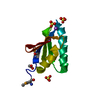
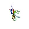
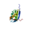
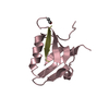
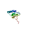
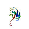
Entry Database : PDB / ID : 2zeqTitle Crystal structure of ubiquitin-like domain of murine Parkin E3 ubiquitin-protein ligase parkin Keywords / / / / / / / / / / / / / / / / / Function / homology Function Domain/homology Component
/ / / / / / / / / / / / / / / / / / / / / / / / / / / / / / / / / / / / / / / / / / / / / / / / / / / / / / / / / / / / / / / / / / / / / / / / / / / / / / / / / / / / / / / / / / / / / / / / / / / / / / / / / / / / / / / / / / / / / / / / / / / / / / / / / / / / / / / / / / / / Biological species Mus musculus (house mouse)Method / / Resolution : 1.65 Å Authors Tomoo, K. Journal : Biochim.Biophys.Acta / Year : 2008Title : Crystal structure and molecular dynamics simulation of ubiquitin-like domain of murine parkinAuthors : Tomoo, K. / Mukai, Y. / In, Y. / Miyagawa, H. / Kitamura, K. / Yamano, A. / Shindo, H. / Ishida, T. History Deposition Dec 14, 2007 Deposition site / Processing site Revision 1.0 Aug 19, 2008 Provider / Type Revision 1.1 Jul 13, 2011 Group Revision 1.2 Mar 13, 2024 Group / Database referencesCategory chem_comp_atom / chem_comp_bond ... chem_comp_atom / chem_comp_bond / database_2 / struct_ref_seq_dif Item / _database_2.pdbx_database_accession / _struct_ref_seq_dif.details
Show all Show less
 Movie
Movie Controller


 Open data
 Basic information
 Components
 Keywords
 Function and homology information
 X-RAY DIFFRACTION /
  Authors
 Citation
 Structure visualization
 Downloads & links
 Other downloads
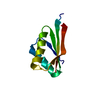


 PDBj
PDBj


 Assembly
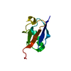

 Mass: 18.015 Da / Num. of mol.: 44 / Source method: isolated from a natural source / Formula: H2O
 Sample preparation
 Processing