 Movie
Movie Controller

[English] 日本語
 Yorodumi
- PDB-1nh9: Crystal Structure of a DNA Binding Protein Mja10b from the hypert... -
+ Open data

- Basic information
| Entry | Database: PDB / ID: 1nh9 | ||||||
|---|---|---|---|---|---|---|---|
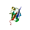
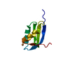
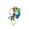
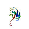
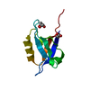
| Title | Crystal Structure of a DNA Binding Protein Mja10b from the hyperthermophile Methanococcus jannaschii | ||||||
 Components Components | DNA-binding protein Alba | ||||||
 Keywords Keywords | DNA BINDING PROTEIN / Mja10b | ||||||
| Function / homology |  Function and homology information Function and homology informationchromosome condensation / chromosome / double-stranded DNA binding / RNA binding / cytoplasm Similarity search - Function | ||||||
| Biological species |   Methanocaldococcus jannaschii (archaea) Methanocaldococcus jannaschii (archaea) | ||||||
| Method |  X-RAY DIFFRACTION / SYNCHROTRON / MOLECULAR REPLACEMENT / Resolution: 2 Å X-RAY DIFFRACTION / SYNCHROTRON / MOLECULAR REPLACEMENT / Resolution: 2 Å | ||||||
 Authors Authors | Wang, G. / Bartlam, M. / Guo, R. / Yang, H. / Xue, H. / Liu, Y. / Huang, L. / Rao, Z. | ||||||
 Citation Citation | Journal: Protein Sci. / Year: 2003 Title: Crystal structure of a DNA binding protein from the hyperthermophilic euryarchaeon Methanococcus jannaschii Authors: Wang, G. / Guo, R. / Bartlam, M. / Yang, H. / Xue, H. / Liu, Y. / Huang, L. / Rao, Z. | ||||||
| History |
|
- Structure visualization
| Structure viewer | Molecule: MolmilJmol/JSmol |
|---|
- Downloads & links
-Download
| PDBx/mmCIF format | 1nh9.cif.gz | 27 KB | Display | PDBx/mmCIF format |
|---|---|---|---|---|
| PDB format | pdb1nh9.ent.gz | 17.8 KB | Display | PDB format |
| PDBx/mmJSON format | 1nh9.json.gz | Tree view | PDBx/mmJSON format | |
| Others |  Other downloads Other downloads |
-Validation report
| Arichive directory | https://data.pdbj.org/pub/pdb/validation_reports/nh/1nh9ftp://data.pdbj.org/pub/pdb/validation_reports/nh/1nh9 | HTTPS FTP |
|---|
-Related structure data
| Related structure data |  1h0xS S: Starting model for refinement |
|---|---|
| Similar structure data |

-Links
 PDBj
PDBj
- Assembly
| Deposited unit | 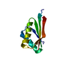
| ||||||||
|---|---|---|---|---|---|---|---|---|---|
| 1 |
| ||||||||
| Unit cell |
| ||||||||
| Details | The biological assembly is a monomer in the asymmetric unit |
-Components
| #1: Protein | Mass: 9690.525 Da / Num. of mol.: 1 Source method: isolated from a genetically manipulated source Source: (gene. exp.) Methanocaldococcus jannaschii (archaea)Plasmid: pET11a / Species (production host): Escherichia coli / Production host:  |
|---|---|
| #2: Water | ChemComp-HOH /  Mass: 18.015 Da / Num. of mol.: 24 / Source method: isolated from a natural source / Formula: H2O Mass: 18.015 Da / Num. of mol.: 24 / Source method: isolated from a natural source / Formula: H2O |
-Experimental details
-Experiment
| Experiment | Method: X-RAY DIFFRACTION / Number of used crystals: 1 |
|---|
- Sample preparation
| Crystal | Density Matthews: 2.73 Å3/Da / Density % sol: 54.69 % | ||||||||||||||||||||||||
|---|---|---|---|---|---|---|---|---|---|---|---|---|---|---|---|---|---|---|---|---|---|---|---|---|---|
| Crystal grow | Temperature: 291 K / Method: vapor diffusion, hanging drop / pH: 8.25 Details: Tris-HCl, MPD, pH 8.25, VAPOR DIFFUSION, HANGING DROP, temperature 291K | ||||||||||||||||||||||||
| Crystal grow | *PLUS Temperature: 291 K / Method: vapor diffusion, hanging drop / Details: Wang, G.G., (2002) Acta Cryst., D58, 1240. | ||||||||||||||||||||||||
| Components of the solutions | *PLUS
|
-Data collection
| Diffraction | Mean temperature: 100 K |
|---|---|
| Diffraction source | Source: SYNCHROTRON / Site: APS  / Beamline: 19-ID / Wavelength: 0.9868 Å / Beamline: 19-ID / Wavelength: 0.9868 Å |
| Detector | Type: SBC-2 / Detector: CCD / Date: Apr 15, 2002 / Details: mirrors |
| Radiation | Monochromator: sagitally focused Si(111) / Protocol: SINGLE WAVELENGTH / Monochromatic (M) / Laue (L): M / Scattering type: x-ray |
| Radiation wavelength | Wavelength: 0.9868 Å / Relative weight: 1 |
| Reflection | Resolution: 2→50 Å / Num. all: 7093 / Num. obs: 7093 / % possible obs: 97.7 % / Observed criterion σ(I): -3 / Redundancy: 13.2 % / Biso Wilson estimate: 18.9 Å2 / Rmerge(I) obs: 0.068 / Net I/σ(I): 21.4 |
| Reflection shell | Resolution: 2→2.07 Å / Redundancy: 13.6 % / Rmerge(I) obs: 0.438 / Mean I/σ(I) obs: 4.2 / Num. unique all: 692 / % possible all: 99.1 |
| Reflection | *PLUS Num. obs: 6946 / % possible obs: 96.6 % / Redundancy: 13.1 % / Num. measured all: 91002 / Rmerge(I) obs: 0.049 |
| Reflection shell | *PLUS Highest resolution: 2 Å / Lowest resolution: 2.06 Å / % possible obs: 99.1 % / Rmerge(I) obs: 0.311 / Mean I/σ(I) obs: 6.2 |
- Processing
| Software |
| ||||||||||||||||||||
|---|---|---|---|---|---|---|---|---|---|---|---|---|---|---|---|---|---|---|---|---|---|
| Refinement | Method to determine structure: MOLECULAR REPLACEMENT Starting model: PDB ENTRY 1h0x Resolution: 2→20 Å / Rfactor Rfree error: 0.006 / Isotropic thermal model: overall / Cross valid method: THROUGHOUT / σ(F): 0 / Stereochemistry target values: Engh & Huber
| ||||||||||||||||||||
| Displacement parameters | Biso mean: 40.2 Å2
| ||||||||||||||||||||
| Refine analyze |
| ||||||||||||||||||||
| Refinement step | Cycle: LAST / Resolution: 2→20 Å
| ||||||||||||||||||||
| Refine LS restraints |
| ||||||||||||||||||||
| LS refinement shell | Resolution: 2→2.06 Å / Rfactor Rfree error: 0.03 / Total num. of bins used: 12
| ||||||||||||||||||||
| Refinement | *PLUS Lowest resolution: 20 Å / % reflection Rfree: 10 % | ||||||||||||||||||||
| Solvent computation | *PLUS | ||||||||||||||||||||
| Displacement parameters | *PLUS | ||||||||||||||||||||
| Refine LS restraints | *PLUS
|
