 Movie
Movie Controller

[English] 日本語
 Yorodumi
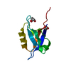
- PDB-6qb2: Crystal structure of the cystatin-based engineered protein scaffo... -
+ Open data

- Basic information
| Entry | Database: PDB / ID: 6qb2 | ||||||
|---|---|---|---|---|---|---|---|
| Title | Crystal structure of the cystatin-based engineered protein scaffold SQT-1C | ||||||
 Components Components | Monomer of SQT-1C | ||||||
 Keywords Keywords | DE NOVO PROTEIN / Engineered Scaffold protein | ||||||
| Function / homology | Nuclear Transport Factor 2; Chain: A, - #10 / Nuclear Transport Factor 2; Chain: A, / Roll / Alpha Beta Function and homology information Function and homology information | ||||||
| Biological species | synthetic construct (others) | ||||||
| Method |  X-RAY DIFFRACTION / SYNCHROTRON / MOLECULAR REPLACEMENT / Resolution: 2.5 Å X-RAY DIFFRACTION / SYNCHROTRON / MOLECULAR REPLACEMENT / Resolution: 2.5 Å | ||||||
 Authors Authors | Levy, C.W. | ||||||
| Funding support |  United Kingdom, 1items United Kingdom, 1items
| ||||||
 Citation Citation | Journal: Sci Rep / Year: 2019 Title: Studies of the oligomerisation mechanism of a cystatin-based engineered protein scaffold. Authors: Zalar, M. / Indrakumar, S. / Levy, C.W. / Tunnicliffe, R.B. / Peters, G.H.J. / Golovanov, A.P. | ||||||
| History |
|
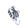
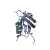
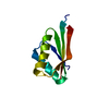
- Structure visualization
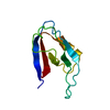
| Structure viewer | Molecule: MolmilJmol/JSmol |
|---|
- Downloads & links
-Download
| PDBx/mmCIF format | 6qb2.cif.gz | 60.1 KB | Display | PDBx/mmCIF format |
|---|---|---|---|---|
| PDB format | pdb6qb2.ent.gz | 37.2 KB | Display | PDB format |
| PDBx/mmJSON format | 6qb2.json.gz | Tree view | PDBx/mmJSON format | |
| Others |  Other downloads Other downloads |
-Validation report
| Arichive directory | https://data.pdbj.org/pub/pdb/validation_reports/qb/6qb2ftp://data.pdbj.org/pub/pdb/validation_reports/qb/6qb2 | HTTPS FTP |
|---|
-Related structure data
| Related structure data | 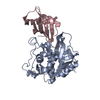 3k9mS S: Starting model for refinement |
|---|---|
| Similar structure data |


-Links
 PDBj
PDBj
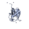
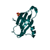
- Assembly
| Deposited unit | 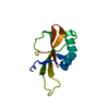
| ||||||||||||
|---|---|---|---|---|---|---|---|---|---|---|---|---|---|
| 1 |
| ||||||||||||
| Unit cell |
|
-Components
| #1: Protein | Mass: 15321.174 Da / Num. of mol.: 1 Source method: isolated from a genetically manipulated source Source: (gene. exp.) synthetic construct (others) / Production host:  |
|---|
-Experimental details
-Experiment
| Experiment | Method: X-RAY DIFFRACTION / Number of used crystals: 1 |
|---|
- Sample preparation
| Crystal | Density Matthews: 3.12 Å3/Da / Density % sol: 60.53 % |
|---|---|
| Crystal grow | Temperature: 277 K / Method: vapor diffusion, sitting drop / Details: 38% Dioxane / Temp details: cold room |
-Data collection
| Diffraction | Mean temperature: 100 K / Serial crystal experiment: N |
|---|---|
| Diffraction source | Source: SYNCHROTRON / Site: Diamond / Beamline: I03 / Wavelength: 0.9 Å |
| Detector | Type: DECTRIS PILATUS 6M / Detector: PIXEL / Date: Apr 22, 2018 |
| Radiation | Protocol: SINGLE WAVELENGTH / Monochromatic (M) / Laue (L): M / Scattering type: x-ray |
| Radiation wavelength | Wavelength: 0.9 Å / Relative weight: 1 |
| Reflection | Resolution: 2.5→42.99 Å / Num. obs: 5209 / % possible obs: 99.79 % / Redundancy: 11.9 % / Biso Wilson estimate: 84.09 Å2 / CC1/2: 0.998 / Rmerge(I) obs: 0.058 / Rpim(I) all: 0.018 / Rrim(I) all: 0.061 / Net I/σ(I): 17.39 |
| Reflection shell | Resolution: 2.5→2.589 Å / Redundancy: 12.6 % / Rmerge(I) obs: 0.92 / Mean I/σ(I) obs: 2.37 / Num. unique obs: 497 / CC1/2: 0.685 / Rpim(I) all: 0.27 / % possible all: 99.6 |
- Processing
| Software |
| ||||||||||||||||||||||||||||||||||||||||
|---|---|---|---|---|---|---|---|---|---|---|---|---|---|---|---|---|---|---|---|---|---|---|---|---|---|---|---|---|---|---|---|---|---|---|---|---|---|---|---|---|---|
| Refinement | Method to determine structure: MOLECULAR REPLACEMENT Starting model: 3K9M Resolution: 2.5→42.99 Å / SU ML: 0.3765 / Cross valid method: FREE R-VALUE / σ(F): 1.5 / Phase error: 28.5026
| ||||||||||||||||||||||||||||||||||||||||
| Solvent computation | Shrinkage radii: 0.9 Å / VDW probe radii: 1.11 Å | ||||||||||||||||||||||||||||||||||||||||
| Displacement parameters | Biso mean: 106.4 Å2 | ||||||||||||||||||||||||||||||||||||||||
| Refinement step | Cycle: LAST / Resolution: 2.5→42.99 Å
| ||||||||||||||||||||||||||||||||||||||||
| Refine LS restraints |
| ||||||||||||||||||||||||||||||||||||||||
| LS refinement shell |
| ||||||||||||||||||||||||||||||||||||||||
| Refinement TLS params. | Method: refined / Origin x: 46.0228387245 Å / Origin y: 21.4256424397 Å / Origin z: 0.780314274909 Å
| ||||||||||||||||||||||||||||||||||||||||
| Refinement TLS group | Selection details: all |
