 Movie
Movie Controller

[English] 日本語
 Yorodumi
- PDB-1h0x: Structure of Alba: an archaeal chromatin protein modulated by ace... -
+ Open data

- Basic information
| Entry | Database: PDB / ID: 1h0x | ||||||
|---|---|---|---|---|---|---|---|
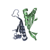
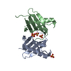
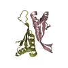
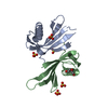
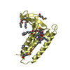
| Title | Structure of Alba: an archaeal chromatin protein modulated by acetylation | ||||||
 Components Components | DNA BINDING PROTEIN SSO10B | ||||||
 Keywords Keywords | DNA BINDING / ARCHAEA / CHROMATIN / ALBA / ACETYLATION | ||||||
| Function / homology |  Function and homology information Function and homology informationnuclease activity / chromosome condensation / chromosome / double-stranded DNA binding / Hydrolases; Acting on ester bonds / hydrolase activity / RNA binding / identical protein binding / cytoplasm Similarity search - Function | ||||||
| Biological species |   SULFOLOBUS SOLFATARICUS (archaea) SULFOLOBUS SOLFATARICUS (archaea) | ||||||
| Method |  X-RAY DIFFRACTION / MAD / Resolution: 2.6 Å X-RAY DIFFRACTION / MAD / Resolution: 2.6 Å | ||||||
 Authors Authors | Wardleworth, B.N. / Russell, R.J.M. / Bell, S.D. / Taylor, G.L. / White, M.F. | ||||||
 Citation Citation | Journal: Embo J. / Year: 2002 Title: Structure of Alba: An Archaeal Chromatin Protein Modulated by Acetylation Authors: Wardleworth, B.N. / Russell, R.J.M. / Bell, S.D. / Taylor, G.L. / White, M.F. #1: Journal: Science / Year: 2002 Title: The Interaction of Alba, a Conserved Archaeal Chromatin Protein, with Sir2 and its Regulation by Acetylation Authors: Bell, S.D. / Botting, C.H. / Wardleworth, B.N. / Jackson, S.P. / White, M.F. #2: Journal: Acta Crystallogr.,Sect.D / Year: 2001 Title: Preliminary Crystallographic Studies of the Double-Stranded DNA Binding Protein Sso10B from Sulfolobus Solfataricus Authors: Wardleworth, B.N. / Russell, R.J.M. / White, M.F. / Taylor, G.L. | ||||||
| History |
|
- Structure visualization
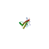
| Structure viewer | Molecule: MolmilJmol/JSmol |
|---|
- Downloads & links
-Download
| PDBx/mmCIF format | 1h0x.cif.gz | 46.7 KB | Display | PDBx/mmCIF format |
|---|---|---|---|---|
| PDB format | pdb1h0x.ent.gz | 34.6 KB | Display | PDB format |
| PDBx/mmJSON format | 1h0x.json.gz | Tree view | PDBx/mmJSON format | |
| Others |  Other downloads Other downloads |
-Validation report
| Arichive directory | https://data.pdbj.org/pub/pdb/validation_reports/h0/1h0xftp://data.pdbj.org/pub/pdb/validation_reports/h0/1h0x | HTTPS FTP |
|---|
-Related structure data


-Links
 PDBj
PDBj

- Assembly
| Deposited unit | 
| ||||||||
|---|---|---|---|---|---|---|---|---|---|
| 1 |
| ||||||||
| Unit cell |
| ||||||||
| Noncrystallographic symmetry (NCS) | NCS oper: (Code: given Matrix: (-0.309, 0.413, 0.857), Vector: |
-Components
| #1: Protein | Mass: 10990.932 Da / Num. of mol.: 2 Source method: isolated from a genetically manipulated source Source: (gene. exp.) SULFOLOBUS SOLFATARICUS (archaea) / Production host:  #2: Water | ChemComp-HOH / |  Mass: 18.015 Da / Num. of mol.: 86 / Source method: isolated from a natural source / Formula: H2O Mass: 18.015 Da / Num. of mol.: 86 / Source method: isolated from a natural source / Formula: H2O |
|---|
-Experimental details
-Experiment
| Experiment | Method: X-RAY DIFFRACTION / Number of used crystals: 1 |
|---|
- Sample preparation
| Crystal | Density Matthews: 4.16 Å3/Da / Density % sol: 70 % | ||||||||||||||||||||||||||||||||||||||||||||||||||||||||
|---|---|---|---|---|---|---|---|---|---|---|---|---|---|---|---|---|---|---|---|---|---|---|---|---|---|---|---|---|---|---|---|---|---|---|---|---|---|---|---|---|---|---|---|---|---|---|---|---|---|---|---|---|---|---|---|---|---|
| Crystal grow | pH: 6.5 / Details: pH 6.50 | ||||||||||||||||||||||||||||||||||||||||||||||||||||||||
| Crystal grow | *PLUS Method: vapor diffusion, hanging dropDetails: Wardleworth, B.N., (2001) Acta Crystallogr.,Sect.D, 57, 1893. | ||||||||||||||||||||||||||||||||||||||||||||||||||||||||
| Components of the solutions | *PLUS
|
-Data collection
| Diffraction | Mean temperature: 100 K |
|---|---|
| Diffraction source | Source: ROTATING ANODE / Type: RIGAKU RU200 / Wavelength: 1.5418 |
| Detector | Type: MSC / Detector: AREA DETECTOR / Date: Jan 15, 2001 / Details: CONFOCAL |
| Radiation | Protocol: SINGLE WAVELENGTH / Monochromatic (M) / Laue (L): M / Scattering type: x-ray |
| Radiation wavelength | Wavelength: 1.5418 Å / Relative weight: 1 |
| Reflection | Resolution: 2.6→30 Å / Num. obs: 10996 / % possible obs: 98 % / Redundancy: 7.6 % / Rmerge(I) obs: 0.046 / Net I/σ(I): 6.9 |
| Reflection shell | Resolution: 2.6→2.69 Å / Redundancy: 7.6 % / Rmerge(I) obs: 0.32 / Mean I/σ(I) obs: 2.7 / % possible all: 95 |
| Reflection | *PLUS Lowest resolution: 30 Å / % possible obs: 98 % |
| Reflection shell | *PLUS % possible obs: 94.7 % |
- Processing
| Software |
| ||||||||||||||||||||||||||||||||||||||||||||||||||||||||||||
|---|---|---|---|---|---|---|---|---|---|---|---|---|---|---|---|---|---|---|---|---|---|---|---|---|---|---|---|---|---|---|---|---|---|---|---|---|---|---|---|---|---|---|---|---|---|---|---|---|---|---|---|---|---|---|---|---|---|---|---|---|---|
| Refinement | Method to determine structure: MAD / Resolution: 2.6→30 Å / Cross valid method: THROUGHOUT
| ||||||||||||||||||||||||||||||||||||||||||||||||||||||||||||
| Refinement step | Cycle: LAST / Resolution: 2.6→30 Å
| ||||||||||||||||||||||||||||||||||||||||||||||||||||||||||||
| Refine LS restraints |
| ||||||||||||||||||||||||||||||||||||||||||||||||||||||||||||
| Refinement | *PLUS Lowest resolution: 30 Å | ||||||||||||||||||||||||||||||||||||||||||||||||||||||||||||
| Solvent computation | *PLUS | ||||||||||||||||||||||||||||||||||||||||||||||||||||||||||||
| Displacement parameters | *PLUS |
