Protocol: SINGLE WAVELENGTH / Monochromatic (M) / Laue (L): M / Scattering type: x-ray
Radiation wavelength
Wavelength: 0.9795 Å / Relative weight: 1
Reflection
Resolution: 1.9→44.9 Å / Num. obs: 40524 / % possible obs: 99.4 % / Observed criterion σ(I): 2 / Redundancy: 13 % / Biso Wilson estimate: 22.59 Å2 / Rmerge(I) obs: 0.07 / Net I/σ(I): 26
Reflection shell
Resolution: 1.92→1.99 Å / Redundancy: 5.8 % / Rmerge(I) obs: 0.93 / Mean I/σ(I) obs: 0.95 / % possible all: 89.3
-
Processing
Software
Name
Version
Classification
PHENIX
(PHENIX.REFINE)
refinement
HKL-2000
datareduction
SCALEPACK
datascaling
SOLVE
phasing
Refinement
Method to determine structure: SAD Starting model: NONE Resolution: 1.92→44.977 Å / SU ML: 0.25 / σ(F): 0.14 / Phase error: 20.85 / Stereochemistry target values: ML Details: THIS MODEL WAS REFINED WITH THE ANOMALOUS DATA (F+ AND F-).
Rfactor
Num. reflection
% reflection
Rfree
0.2276
3579
5.1 %
Rwork
0.1738
-
-
obs
0.1764
40524
91.33 %
Solvent computation
Shrinkage radii: 0.9 Å / VDW probe radii: 1.11 Å / Solvent model: FLAT BULK SOLVENT MODEL / Bsol: 49.659 Å2 / ksol: 0.383 e/Å3
Displacement parameters
Biso mean: 30.58 Å2
Baniso -1
Baniso -2
Baniso -3
1-
-2.5949 Å2
0 Å2
-0 Å2
2-
-
2.9559 Å2
0 Å2
3-
-
-
-2.8033 Å2
Refinement step
Cycle: LAST / Resolution: 1.92→44.977 Å
Protein
Nucleic acid
Ligand
Solvent
Total
Num. atoms
3371
0
18
290
3679
Refine LS restraints
Refine-ID
Type
Dev ideal
Number
X-RAY DIFFRACTION
f_bond_d
0.011
3464
X-RAY DIFFRACTION
f_angle_d
1.294
4707
X-RAY DIFFRACTION
f_dihedral_angle_d
16.866
1253
X-RAY DIFFRACTION
f_chiral_restr
0.084
546
X-RAY DIFFRACTION
f_plane_restr
0.005
618
LS refinement shell
Resolution (Å)
Rfactor Rfree
Num. reflection Rfree
Rfactor Rwork
Num. reflection Rwork
Refine-ID
% reflection obs (%)
1.9182-1.9434
0.3034
78
0.3212
1747
X-RAY DIFFRACTION
62
1.9434-1.97
0.2906
104
0.2737
2198
X-RAY DIFFRACTION
77
1.97-1.9982
0.2622
126
0.2505
2223
X-RAY DIFFRACTION
79
1.9982-2.028
0.2755
132
0.2291
2313
X-RAY DIFFRACTION
83
2.028-2.0597
0.2552
113
0.1831
2348
X-RAY DIFFRACTION
82
2.0597-2.0935
0.2223
116
0.1992
2485
X-RAY DIFFRACTION
88
2.0935-2.1296
0.2033
153
0.1781
2598
X-RAY DIFFRACTION
91
2.1296-2.1683
0.2119
162
0.1762
2562
X-RAY DIFFRACTION
92
2.1683-2.21
0.2842
153
0.1942
2550
X-RAY DIFFRACTION
92
2.21-2.2551
0.278
139
0.2263
2466
X-RAY DIFFRACTION
87
2.2551-2.3041
0.2689
131
0.1877
2375
X-RAY DIFFRACTION
85
2.3041-2.3577
0.2187
136
0.1664
2668
X-RAY DIFFRACTION
95
2.3577-2.4167
0.2395
145
0.1709
2695
X-RAY DIFFRACTION
95
2.4167-2.482
0.2522
140
0.167
2652
X-RAY DIFFRACTION
94
2.482-2.555
0.2013
175
0.1633
2646
X-RAY DIFFRACTION
95
2.555-2.6375
0.2154
143
0.1628
2741
X-RAY DIFFRACTION
96
2.6375-2.7318
0.272
163
0.1642
2687
X-RAY DIFFRACTION
96
2.7318-2.8411
0.2213
135
0.1644
2767
X-RAY DIFFRACTION
97
2.8411-2.9704
0.244
154
0.1605
2721
X-RAY DIFFRACTION
97
2.9704-3.127
0.2164
142
0.1596
2766
X-RAY DIFFRACTION
98
3.127-3.3228
0.1811
151
0.1505
2767
X-RAY DIFFRACTION
99
3.3228-3.5793
0.1859
142
0.1437
2806
X-RAY DIFFRACTION
98
3.5793-3.9393
0.2168
144
0.1553
2784
X-RAY DIFFRACTION
99
3.9393-4.5089
0.2235
117
0.1474
2832
X-RAY DIFFRACTION
99
4.5089-5.6789
0.1947
136
0.1658
2829
X-RAY DIFFRACTION
100
5.6789-44.9891
0.2158
149
0.1936
2807
X-RAY DIFFRACTION
100
Refinement TLS params.
Method: refined / Refine-ID: X-RAY DIFFRACTION
ID
L11 (°2)
L12 (°2)
L13 (°2)
L22 (°2)
L23 (°2)
L33 (°2)
S11 (Å °)
S12 (Å °)
S13 (Å °)
S21 (Å °)
S22 (Å °)
S23 (Å °)
S31 (Å °)
S32 (Å °)
S33 (Å °)
T11 (Å2)
T12 (Å2)
T13 (Å2)
T22 (Å2)
T23 (Å2)
T33 (Å2)
Origin x (Å)
Origin y (Å)
Origin z (Å)
1
0.8428
-0.2131
0.572
0.5207
-0.5484
1.1595
-0.0545
0.0188
0.0086
0.0326
-0.0042
0.0445
-0.1272
0.0478
0.0476
0.1736
0.0064
-0.0077
0.1405
0.01
0.1593
58.5611
47.5885
33.9546
2
1.1895
-0.2517
0.0025
2.698
1.1252
0.1488
-0.1788
-0.4097
0.1563
0.2953
0.1248
0.1324
-0.0368
-0.0431
0.0609
0.2679
0.0708
-0.0093
0.2656
-0.034
0.1792
55.3528
56.3941
51.7073
3
1.2365
-0.2923
-0.0376
1.2035
-0.3094
0.8179
-0.0188
-0.0834
0.0079
0.0611
-0.0722
-0.0977
0.0126
0.181
0.0878
0.1078
0.0087
-0.0111
0.1489
0.0285
0.1268
70.3426
17.3566
57.8791
Refinement TLS group
ID
Refine-ID
Refine TLS-ID
Selection details
1
X-RAY DIFFRACTION
1
CHAINAANDRESID186:326
2
X-RAY DIFFRACTION
2
CHAINAANDRESID327:445
3
X-RAY DIFFRACTION
3
CHAINAANDRESID446:627
+
About Yorodumi
-
News
-
Feb 9, 2022. New format data for meta-information of EMDB entries
New format data for meta-information of EMDB entries
Version 3 of the EMDB header file is now the official format.
The previous official version 1.9 will be removed from the archive.
In the structure databanks used in Yorodumi, some data are registered as the other names, "COVID-19 virus" and "2019-nCoV". Here are the details of the virus and the list of structure data.
Jan 31, 2019. EMDB accession codes are about to change! (news from PDBe EMDB page)
EMDB accession codes are about to change! (news from PDBe EMDB page)
The allocation of 4 digits for EMDB accession codes will soon come to an end. Whilst these codes will remain in use, new EMDB accession codes will include an additional digit and will expand incrementally as the available range of codes is exhausted. The current 4-digit format prefixed with “EMD-” (i.e. EMD-XXXX) will advance to a 5-digit format (i.e. EMD-XXXXX), and so on. It is currently estimated that the 4-digit codes will be depleted around Spring 2019, at which point the 5-digit format will come into force.
The EM Navigator/Yorodumi systems omit the EMD- prefix.
Related info.:Q: What is EMD? / ID/Accession-code notation in Yorodumi/EM Navigator
Yorodumi is a browser for structure data from EMDB, PDB, SASBDB, etc.
This page is also the successor to EM Navigator detail page, and also detail information page/front-end page for Omokage search.
The word "yorodu" (or yorozu) is an old Japanese word meaning "ten thousand". "mi" (miru) is to see.
Related info.:EMDB / PDB / SASBDB / Comparison of 3 databanks / Yorodumi Search / Aug 31, 2016. New EM Navigator & Yorodumi / Yorodumi Papers / Jmol/JSmol / Function and homology information / Changes in new EM Navigator and Yorodumi
 Movie
Movie Controller

 Yorodumi
 Open data
 Basic information
 Components
 Keywords
 Function and homology information
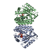
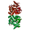
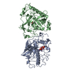
 STREPTOCOCCUS PNEUMONIAE (bacteria)
 X-RAY DIFFRACTION /
  Authors
 Citation
 Structure visualization
 Downloads & links
 Other downloads
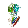


 PDBj
PDBj Assembly

 Mass: 92.094 Da / Num. of mol.: 3 / Source method: obtained synthetically / Formula: C3H8O3
 Mass: 18.015 Da / Num. of mol.: 290 / Source method: isolated from a natural source / Formula: H2O
 Sample preparation
 / Beamline: 5.0.2 / Wavelength: 0.9795
  Processing