 Movie
Movie Controller

[English] 日本語
 Yorodumi
- PDB-6iu8: Crystal structure of cytoplasmic metal binding domain with cobalt ions -
+ Open data

- Basic information
| Entry | Database: PDB / ID: 6iu8 | ||||||
|---|---|---|---|---|---|---|---|
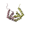
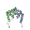
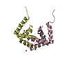
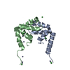
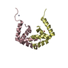
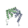
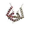
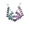
| Title | Crystal structure of cytoplasmic metal binding domain with cobalt ions | ||||||
 Components Components | VIT1 | ||||||
 Keywords Keywords | METAL TRANSPORT / membrane protein | ||||||
| Function / homology | :  Function and homology information Function and homology information | ||||||
| Biological species |  Eucalyptus grandis (rose gum) Eucalyptus grandis (rose gum) | ||||||
| Method |  X-RAY DIFFRACTION / SYNCHROTRON / MOLECULAR REPLACEMENT / Resolution: 2.7 Å X-RAY DIFFRACTION / SYNCHROTRON / MOLECULAR REPLACEMENT / Resolution: 2.7 Å | ||||||
 Authors Authors | Kato, T. / Nishizawa, T. / Yamashita, K. / Kumazaki, K. / Ishitani, R. / Nureki, O. | ||||||
 Citation Citation | Journal: Nat Plants / Year: 2019 Title: Crystal structure of plant vacuolar iron transporter VIT1. Authors: Kato, T. / Kumazaki, K. / Wada, M. / Taniguchi, R. / Nakane, T. / Yamashita, K. / Hirata, K. / Ishitani, R. / Ito, K. / Nishizawa, T. / Nureki, O. | ||||||
| History |
|
- Structure visualization
| Structure viewer | Molecule: MolmilJmol/JSmol |
|---|
- Downloads & links
-Download
| PDBx/mmCIF format | 6iu8.cif.gz | 135.6 KB | Display | PDBx/mmCIF format |
|---|---|---|---|---|
| PDB format | pdb6iu8.ent.gz | 105.2 KB | Display | PDB format |
| PDBx/mmJSON format | 6iu8.json.gz | Tree view | PDBx/mmJSON format | |
| Others |  Other downloads Other downloads |
-Validation report
| Arichive directory | https://data.pdbj.org/pub/pdb/validation_reports/iu/6iu8ftp://data.pdbj.org/pub/pdb/validation_reports/iu/6iu8 | HTTPS FTP |
|---|
-Related structure data
| Related structure data |  6iu3C 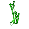 6iu4C  6iu5SC  6iu6C  6iu9C S: Starting model for refinement C: citing same article ( |
|---|---|
| Similar structure data | |
| Experimental dataset #1 | Data reference: 10.5281/zenodo.2532134 / Data set type: diffraction image data |


-Links
 PDBj
PDBj
- Assembly
| Deposited unit | 
| ||||||||
|---|---|---|---|---|---|---|---|---|---|
| 1 | 
| ||||||||
| 2 | 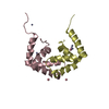
| ||||||||
| 3 | 
| ||||||||
| 4 | 
| ||||||||
| Unit cell |
|
-Components
| #1: Protein | Mass: 9106.312 Da / Num. of mol.: 8 Source method: isolated from a genetically manipulated source Source: (gene. exp.) Eucalyptus grandis (rose gum) / Plasmid: modified pE-SUMO / Production host:  #2: Chemical | ChemComp-ZN /   Mass: 65.409 Da / Num. of mol.: 15 / Source method: obtained synthetically / Formula: Zn Mass: 65.409 Da / Num. of mol.: 15 / Source method: obtained synthetically / Formula: Zn#3: Chemical | ChemComp-CO /   Mass: 58.933 Da / Num. of mol.: 18 / Source method: obtained synthetically / Formula: Co Mass: 58.933 Da / Num. of mol.: 18 / Source method: obtained synthetically / Formula: Co#4: Water | ChemComp-HOH / |  Mass: 18.015 Da / Num. of mol.: 8 / Source method: isolated from a natural source / Formula: H2O Mass: 18.015 Da / Num. of mol.: 8 / Source method: isolated from a natural source / Formula: H2O |
|---|
-Experimental details
-Experiment
| Experiment | Method: X-RAY DIFFRACTION / Number of used crystals: 1 |
|---|
- Sample preparation
| Crystal | Density Matthews: 2.85 Å3/Da / Density % sol: 56.83 % / Mosaicity: 0.16 ° |
|---|---|
| Crystal grow | Temperature: 293 K / Method: vapor diffusion, sitting drop / pH: 7 Details: 21-23% PEG600, 0.1 M HEPES pH7.0 and 0.001-0.003 M zinc cloride |
-Data collection
| Diffraction | Mean temperature: 100 K / Serial crystal experiment: N | |||||||||||||||||||||||||||
|---|---|---|---|---|---|---|---|---|---|---|---|---|---|---|---|---|---|---|---|---|---|---|---|---|---|---|---|---|
| Diffraction source | Source: SYNCHROTRON / Site: SPring-8  / Beamline: BL41XU / Wavelength: 1.605 Å / Beamline: BL41XU / Wavelength: 1.605 Å | |||||||||||||||||||||||||||
| Detector | Type: DECTRIS PILATUS3 6M / Detector: PIXEL / Date: Jul 26, 2017 | |||||||||||||||||||||||||||
| Radiation | Monochromator: Si(111) / Protocol: SINGLE WAVELENGTH / Monochromatic (M) / Laue (L): M / Scattering type: x-ray | |||||||||||||||||||||||||||
| Radiation wavelength | Wavelength: 1.605 Å / Relative weight: 1 | |||||||||||||||||||||||||||
| Reflection twin |
| |||||||||||||||||||||||||||
| Reflection | Resolution: 2.7→19.72 Å / Num. obs: 22053 / % possible obs: 99.7 % / Redundancy: 10.4 % / CC1/2: 0.997 / Rmerge(I) obs: 0.131 / Rpim(I) all: 0.042 / Rrim(I) all: 0.138 / Net I/σ(I): 11.2 / Num. measured all: 230049 / Scaling rejects: 54 | |||||||||||||||||||||||||||
| Reflection shell | Diffraction-ID: 1 / Redundancy: 10.5 %
|
- Processing
| Software |
| ||||||||||||||||||||||||||||||||||||||||||||||||||||||||||||
|---|---|---|---|---|---|---|---|---|---|---|---|---|---|---|---|---|---|---|---|---|---|---|---|---|---|---|---|---|---|---|---|---|---|---|---|---|---|---|---|---|---|---|---|---|---|---|---|---|---|---|---|---|---|---|---|---|---|---|---|---|---|
| Refinement | Method to determine structure: MOLECULAR REPLACEMENT Starting model: 6IU5 Resolution: 2.7→19.72 Å / Cor.coef. Fo:Fc: 0.965 / Cor.coef. Fo:Fc free: 0.93 / SU B: 9.915 / SU ML: 0.215 / Cross valid method: THROUGHOUT / σ(F): 0 / ESU R: 0.142 / ESU R Free: 0.059 / Stereochemistry target values: MAXIMUM LIKELIHOOD Details: HYDROGENS HAVE BEEN ADDED IN THE RIDING POSITIONS U VALUES : REFINED INDIVIDUALLY
| ||||||||||||||||||||||||||||||||||||||||||||||||||||||||||||
| Solvent computation | Ion probe radii: 0.8 Å / Shrinkage radii: 0.8 Å / VDW probe radii: 1.2 Å / Solvent model: MASK | ||||||||||||||||||||||||||||||||||||||||||||||||||||||||||||
| Displacement parameters | Biso max: 136.96 Å2 / Biso mean: 66.018 Å2 / Biso min: 24.9 Å2
| ||||||||||||||||||||||||||||||||||||||||||||||||||||||||||||
| Refinement step | Cycle: final / Resolution: 2.7→19.72 Å
| ||||||||||||||||||||||||||||||||||||||||||||||||||||||||||||
| Refine LS restraints |
| ||||||||||||||||||||||||||||||||||||||||||||||||||||||||||||
| LS refinement shell | Resolution: 2.7→2.771 Å / Rfactor Rfree error: 0 / Total num. of bins used: 20
|
