 Movie
Movie Controller


+ Open data

- Basic information
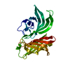
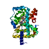
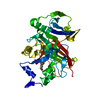
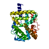
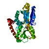
| Entry | Database: PDB / ID: 6in7 | ||||||
|---|---|---|---|---|---|---|---|
| Title | Crystal structure of AlgU in complex with MucA(cyto) | ||||||
 Components Components |
| ||||||
 Keywords Keywords | TRANSCRIPTION / Sigma factor | ||||||
| Function / homology |  Function and homology information Function and homology informationregulation of polysaccharide biosynthetic process / positive regulation of response to oxidative stress / positive regulation of cellular response to heat / positive regulation of cell adhesion involved in single-species biofilm formation / positive regulation of single-species biofilm formation on inanimate substrate / alginic acid biosynthetic process / negative regulation of bacterial-type flagellum-dependent cell motility / sigma factor antagonist activity / DNA-binding transcription activator activity / cellular response to cell envelope stress ...regulation of polysaccharide biosynthetic process / positive regulation of response to oxidative stress / positive regulation of cellular response to heat / positive regulation of cell adhesion involved in single-species biofilm formation / positive regulation of single-species biofilm formation on inanimate substrate / alginic acid biosynthetic process / negative regulation of bacterial-type flagellum-dependent cell motility / sigma factor antagonist activity / DNA-binding transcription activator activity / cellular response to cell envelope stress / cellular response to antibiotic / sigma factor activity / cytosolic DNA-directed RNA polymerase complex / DNA-templated transcription initiation / protein-DNA complex / transcription cis-regulatory region binding / regulation of DNA-templated transcription / positive regulation of DNA-templated transcription / plasma membrane Similarity search - Function | ||||||
| Biological species |   Pseudomonas aeruginosa (bacteria) Pseudomonas aeruginosa (bacteria) | ||||||
| Method |  X-RAY DIFFRACTION / SYNCHROTRON / MOLECULAR REPLACEMENT / Resolution: 1.96 Å X-RAY DIFFRACTION / SYNCHROTRON / MOLECULAR REPLACEMENT / Resolution: 1.96 Å | ||||||
 Authors Authors | Li, S. / Zhang, Q. / Bartlam, M. | ||||||
| Funding support |  China, 1items China, 1items
| ||||||
 Citation Citation | Journal: Febs J. / Year: 2019 Title: Structural basis for the recognition of MucA by MucB and AlgU in Pseudomonas aeruginosa. Authors: Li, S. / Lou, X. / Xu, Y. / Teng, X. / Liu, R. / Zhang, Q. / Wu, W. / Wang, Y. / Bartlam, M. | ||||||
| History |
|
- Structure visualization
| Structure viewer | Molecule: MolmilJmol/JSmol |
|---|
- Downloads & links
-Download
| PDBx/mmCIF format | 6in7.cif.gz | 72.4 KB | Display | PDBx/mmCIF format |
|---|---|---|---|---|
| PDB format | pdb6in7.ent.gz | 52.5 KB | Display | PDB format |
| PDBx/mmJSON format | 6in7.json.gz | Tree view | PDBx/mmJSON format | |
| Others |  Other downloads Other downloads |
-Validation report
| Arichive directory | https://data.pdbj.org/pub/pdb/validation_reports/in/6in7ftp://data.pdbj.org/pub/pdb/validation_reports/in/6in7 | HTTPS FTP |
|---|
-Related structure data
| Related structure data | 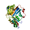 6in8C 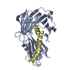 6in9C  1or7S S: Starting model for refinement C: citing same article ( |
|---|---|
| Similar structure data |


-Links
 PDBj
PDBj
- Assembly
| Deposited unit | 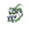
| ||||||||
|---|---|---|---|---|---|---|---|---|---|
| 1 |
| ||||||||
| Unit cell |
|
-Components
| #1: Protein | Mass: 8908.094 Da / Num. of mol.: 1 Source method: isolated from a genetically manipulated source Source: (gene. exp.) Pseudomonas aeruginosa (strain ATCC 15692 / DSM 22644 / CIP 104116 / JCM 14847 / LMG 12228 / 1C / PRS 101 / PAO1) (bacteria)Strain: ATCC 15692 / DSM 22644 / CIP 104116 / JCM 14847 / LMG 12228 / 1C / PRS 101 / PAO1 Gene: mucA, PA0763 / Production host: |
|---|---|
| #2: Protein | Mass: 22227.156 Da / Num. of mol.: 1 Source method: isolated from a genetically manipulated source Source: (gene. exp.) Pseudomonas aeruginosa (strain ATCC 15692 / DSM 22644 / CIP 104116 / JCM 14847 / LMG 12228 / 1C / PRS 101 / PAO1) (bacteria)Strain: ATCC 15692 / DSM 22644 / CIP 104116 / JCM 14847 / LMG 12228 / 1C / PRS 101 / PAO1 Gene: algU, algT, PA0762 / Production host: |
| #3: Chemical | ChemComp-NCA / 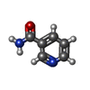  Mass: 122.125 Da / Num. of mol.: 1 / Source method: obtained synthetically / Formula: C6H6N2O / Comment: medication*YM Mass: 122.125 Da / Num. of mol.: 1 / Source method: obtained synthetically / Formula: C6H6N2O / Comment: medication*YM |
| #4: Water | ChemComp-HOH /  Mass: 18.015 Da / Num. of mol.: 327 / Source method: isolated from a natural source / Formula: H2O Mass: 18.015 Da / Num. of mol.: 327 / Source method: isolated from a natural source / Formula: H2O |
-Experimental details
-Experiment
| Experiment | Method: X-RAY DIFFRACTION / Number of used crystals: 1 |
|---|
- Sample preparation
| Crystal | Density Matthews: 2.6 Å3/Da / Density % sol: 52.65 % |
|---|---|
| Crystal grow | Temperature: 293 K / Method: vapor diffusion, sitting drop / pH: 6.5 Details: 2.0 M ammonium sulfate,0.1 M cacodylate pH 6.5, 0.2M sodium chloride |
-Data collection
| Diffraction | Mean temperature: 100 K / Serial crystal experiment: N |
|---|---|
| Diffraction source | Source: SYNCHROTRON / Site: SSRF / Beamline: BL18U1 / Wavelength: 0.977853 Å |
| Detector | Type: DECTRIS PILATUS 6M / Detector: PIXEL / Date: Jun 4, 2017 |
| Radiation | Protocol: SINGLE WAVELENGTH / Monochromatic (M) / Laue (L): M / Scattering type: x-ray |
| Radiation wavelength | Wavelength: 0.977853 Å / Relative weight: 1 |
| Reflection | Resolution: 1.96→50 Å / Num. obs: 23934 / % possible obs: 99.5 % / Redundancy: 6.2 % / Rpim(I) all: 0.09 / Net I/σ(I): 27.9 |
| Reflection shell | Resolution: 1.96→1.99 Å / Redundancy: 5.6 % / Mean I/σ(I) obs: 9.2 / Num. unique obs: 1053 / Rpim(I) all: 0.276 / % possible all: 90.6 |
- Processing
| Software |
| |||||||||||||||||||||||||||||||||||||||||||||||||||||||||||||||
|---|---|---|---|---|---|---|---|---|---|---|---|---|---|---|---|---|---|---|---|---|---|---|---|---|---|---|---|---|---|---|---|---|---|---|---|---|---|---|---|---|---|---|---|---|---|---|---|---|---|---|---|---|---|---|---|---|---|---|---|---|---|---|---|---|
| Refinement | Method to determine structure: MOLECULAR REPLACEMENT Starting model: 1OR7 Resolution: 1.96→45.505 Å / SU ML: 0.15 / Cross valid method: FREE R-VALUE / σ(F): 1.38 / Phase error: 19.04
| |||||||||||||||||||||||||||||||||||||||||||||||||||||||||||||||
| Solvent computation | Shrinkage radii: 0.9 Å / VDW probe radii: 1.11 Å | |||||||||||||||||||||||||||||||||||||||||||||||||||||||||||||||
| Refinement step | Cycle: LAST / Resolution: 1.96→45.505 Å
| |||||||||||||||||||||||||||||||||||||||||||||||||||||||||||||||
| Refine LS restraints |
| |||||||||||||||||||||||||||||||||||||||||||||||||||||||||||||||
| LS refinement shell |
|
