 Movie
Movie Controller

[English] 日本語
 Yorodumi
- PDB-3p3f: Crystal structure of the F36A mutant of the fluoroacetyl-CoA-spec... -
+ Open data

- Basic information
| Entry | Database: PDB / ID: 3p3f | ||||||
|---|---|---|---|---|---|---|---|
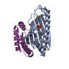
| Title | Crystal structure of the F36A mutant of the fluoroacetyl-CoA-specific thioesterase FlK | ||||||
 Components Components | Fluoroacetyl coenzyme A thioesterase | ||||||
 Keywords Keywords | HYDROLASE / hot dog-fold / thioesterase | ||||||
| Function / homology |  Function and homology information Function and homology informationfluoroacetyl-CoA thioesterase / acyl-CoA hydrolase activity / protein homodimerization activity Similarity search - Function | ||||||
| Biological species |  Streptomyces cattleya (bacteria) Streptomyces cattleya (bacteria) | ||||||
| Method |  X-RAY DIFFRACTION / SYNCHROTRON / MOLECULAR REPLACEMENT / Resolution: 2.3 Å X-RAY DIFFRACTION / SYNCHROTRON / MOLECULAR REPLACEMENT / Resolution: 2.3 Å | ||||||
 Authors Authors | Weeks, A.M. / Coyle, S.M. / Jinek, M. / Doudna, J.A. / Chang, M.C.Y. | ||||||
 Citation Citation | Journal: Biochemistry / Year: 2010 Title: Structural and biochemical studies of a fluoroacetyl-CoA-specific thioesterase reveal a molecular basis for fluorine selectivity. Authors: Weeks, A.M. / Coyle, S.M. / Jinek, M. / Doudna, J.A. / Chang, M.C. | ||||||
| History |
|
- Structure visualization
| Structure viewer | Molecule: MolmilJmol/JSmol |
|---|
- Downloads & links
-Download
| PDBx/mmCIF format | 3p3f.cif.gz | 319.9 KB | Display | PDBx/mmCIF format |
|---|---|---|---|---|
| PDB format | pdb3p3f.ent.gz | 261.6 KB | Display | PDB format |
| PDBx/mmJSON format | 3p3f.json.gz | Tree view | PDBx/mmJSON format | |
| Others |  Other downloads Other downloads |
-Validation report
| Arichive directory | https://data.pdbj.org/pub/pdb/validation_reports/p3/3p3fftp://data.pdbj.org/pub/pdb/validation_reports/p3/3p3f | HTTPS FTP |
|---|
-Related structure data


-Links
 PDBj
PDBj
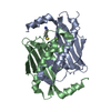
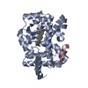
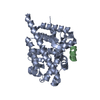
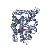
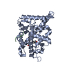
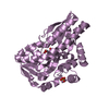
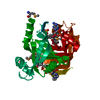
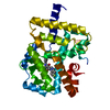
- Assembly
| Deposited unit | 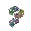
| |||||||||||||||
|---|---|---|---|---|---|---|---|---|---|---|---|---|---|---|---|---|
| 1 | 
| |||||||||||||||
| 2 | 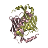
| |||||||||||||||
| 3 | 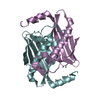
| |||||||||||||||
| Unit cell |
| |||||||||||||||
| Components on special symmetry positions |
|
-Components
| #1: Protein | Mass: 15489.625 Da / Num. of mol.: 6 / Mutation: F36A Source method: isolated from a genetically manipulated source Source: (gene. exp.) Streptomyces cattleya (bacteria) / Gene: flK / Production host: #2: Water | ChemComp-HOH / |  Mass: 18.015 Da / Num. of mol.: 559 / Source method: isolated from a natural source / Formula: H2O Mass: 18.015 Da / Num. of mol.: 559 / Source method: isolated from a natural source / Formula: H2OHas protein modification | Y | |
|---|
-Experimental details
-Experiment
| Experiment | Method: X-RAY DIFFRACTION / Number of used crystals: 1 |
|---|
- Sample preparation
| Crystal | Density Matthews: 2.12 Å3/Da / Density % sol: 42 % |
|---|---|
| Crystal grow | Temperature: 291 K / Method: vapor diffusion, hanging drop / pH: 7.6 Details: 0.1 M Tris-HCl, pH 7.6 25% PEG 3350, VAPOR DIFFUSION, HANGING DROP, temperature 291K |
-Data collection
| Diffraction | Mean temperature: 100 K |
|---|---|
| Diffraction source | Source: SYNCHROTRON / Site: ALS  / Beamline: 8.3.1 / Wavelength: 1.116 Å / Beamline: 8.3.1 / Wavelength: 1.116 Å |
| Detector | Type: ADSC QUANTUM 315r / Detector: CCD / Date: May 7, 2010 |
| Radiation | Monochromator: Double flat crystal Si(111) / Protocol: SINGLE WAVELENGTH / Monochromatic (M) / Laue (L): M / Scattering type: x-ray |
| Radiation wavelength | Wavelength: 1.116 Å / Relative weight: 1 |
| Reflection | Resolution: 2.3→19.75 Å / Num. all: 34432 / Num. obs: 34432 / % possible obs: 99.7 % / Observed criterion σ(F): -3 / Observed criterion σ(I): 0 |
| Reflection shell | Resolution: 2.3→2.48 Å / % possible all: 99.7 |
- Processing
| Software |
| |||||||||||||||||||||||||||||||||||||||||||||||||||||||||||||||||||||||||||||||||||||||||||
|---|---|---|---|---|---|---|---|---|---|---|---|---|---|---|---|---|---|---|---|---|---|---|---|---|---|---|---|---|---|---|---|---|---|---|---|---|---|---|---|---|---|---|---|---|---|---|---|---|---|---|---|---|---|---|---|---|---|---|---|---|---|---|---|---|---|---|---|---|---|---|---|---|---|---|---|---|---|---|---|---|---|---|---|---|---|---|---|---|---|---|---|---|
| Refinement | Method to determine structure: MOLECULAR REPLACEMENT / Resolution: 2.3→19.75 Å / SU ML: 0.33 / σ(F): 1.99 / Phase error: 24.13 / Stereochemistry target values: ML
| |||||||||||||||||||||||||||||||||||||||||||||||||||||||||||||||||||||||||||||||||||||||||||
| Solvent computation | Shrinkage radii: 0.9 Å / VDW probe radii: 1.11 Å / Solvent model: FLAT BULK SOLVENT MODEL / Bsol: 63.131 Å2 / ksol: 0.407 e/Å3 | |||||||||||||||||||||||||||||||||||||||||||||||||||||||||||||||||||||||||||||||||||||||||||
| Displacement parameters |
| |||||||||||||||||||||||||||||||||||||||||||||||||||||||||||||||||||||||||||||||||||||||||||
| Refinement step | Cycle: LAST / Resolution: 2.3→19.75 Å
| |||||||||||||||||||||||||||||||||||||||||||||||||||||||||||||||||||||||||||||||||||||||||||
| Refine LS restraints |
| |||||||||||||||||||||||||||||||||||||||||||||||||||||||||||||||||||||||||||||||||||||||||||
| LS refinement shell |
|
