 Movie
Movie Controller

[English] 日本語
 Yorodumi
- PDB-2pfz: Crystal structure of DctP6, a Bordetella pertussis extracytoplasm... -
+ Open data

- Basic information
| Entry | Database: PDB / ID: 2pfz | ||||||
|---|---|---|---|---|---|---|---|
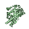
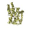
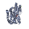
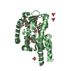
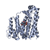
| Title | Crystal structure of DctP6, a Bordetella pertussis extracytoplasmic solute receptor binding pyroglutamic acid | ||||||
 Components Components | Putative exported protein | ||||||
 Keywords Keywords | TRANSPORT PROTEIN / extracytoplasmic solute receptor / tripartite ATP independent periplasmic transport / pyroglutamic acid / ligand binding | ||||||
| Function / homology |  Function and homology information Function and homology information | ||||||
| Biological species |  Bordetella pertussis Tohama I (bacteria) Bordetella pertussis Tohama I (bacteria) | ||||||
| Method |  X-RAY DIFFRACTION / SYNCHROTRON / MOLECULAR REPLACEMENT / Resolution: 1.8 Å X-RAY DIFFRACTION / SYNCHROTRON / MOLECULAR REPLACEMENT / Resolution: 1.8 Å | ||||||
 Authors Authors | Rucktooa, P. | ||||||
 Citation Citation | Journal: J.Mol.Biol. / Year: 2007 Title: Crystal Structures of two Bordetella pertussis Periplasmic Receptors Contribute to Defining a Novel Pyroglutamic Acid Binding DctP Subfamily. Authors: Rucktooa, P. / Antoine, R. / Herrou, J. / Huvent, I. / Locht, C. / Jacob-Dubuisson, F. / Villeret, V. / Bompard, C. | ||||||
| History |
|
- Structure visualization
| Structure viewer | Molecule: MolmilJmol/JSmol |
|---|
- Downloads & links
-Download
| PDBx/mmCIF format | 2pfz.cif.gz | 131.5 KB | Display | PDBx/mmCIF format |
|---|---|---|---|---|
| PDB format | pdb2pfz.ent.gz | 103.6 KB | Display | PDB format |
| PDBx/mmJSON format | 2pfz.json.gz | Tree view | PDBx/mmJSON format | |
| Others |  Other downloads Other downloads |
-Validation report
| Arichive directory | https://data.pdbj.org/pub/pdb/validation_reports/pf/2pfzftp://data.pdbj.org/pub/pdb/validation_reports/pf/2pfz | HTTPS FTP |
|---|
-Related structure data


-Links
 PDBj
PDBj- Assembly
| Deposited unit | 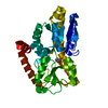
| ||||||||
|---|---|---|---|---|---|---|---|---|---|
| 1 |
| ||||||||
| Unit cell |
| ||||||||
| Components on special symmetry positions |
|
-Components
| #1: Protein | Mass: 33350.043 Da / Num. of mol.: 1 Source method: isolated from a genetically manipulated source Source: (gene. exp.) Bordetella pertussis Tohama I (bacteria)Species: Bordetella pertussis / Strain: Tohama I / Gene: bp1887 / Plasmid: pQE30 / Species (production host): Escherichia coli / Production host: |
|---|---|
| #2: Chemical | ChemComp-PCA /   Type: L-peptide linking / Mass: 129.114 Da / Num. of mol.: 1 / Source method: obtained synthetically / Formula: C5H7NO3 Type: L-peptide linking / Mass: 129.114 Da / Num. of mol.: 1 / Source method: obtained synthetically / Formula: C5H7NO3 |
| #3: Water | ChemComp-HOH /  Mass: 18.015 Da / Num. of mol.: 181 / Source method: isolated from a natural source / Formula: H2O Mass: 18.015 Da / Num. of mol.: 181 / Source method: isolated from a natural source / Formula: H2O |
-Experimental details
-Experiment
| Experiment | Method: X-RAY DIFFRACTION / Number of used crystals: 1 |
|---|
- Sample preparation
| Crystal | Density Matthews: 2.78 Å3/Da / Density % sol: 55.71 % |
|---|---|
| Crystal grow | Temperature: 277 K / Method: vapor diffusion, hanging drop / pH: 6.5 Details: 36% (w/v) PEG 2000 MME, 0.1M PIPES, pH 6.5, VAPOR DIFFUSION, HANGING DROP, temperature 277K |
-Data collection
| Diffraction source | Source: SYNCHROTRON / Site: ESRF  / Beamline: BM30A / Wavelength: 0.97942 Å / Beamline: BM30A / Wavelength: 0.97942 Å |
|---|---|
| Detector | Type: MAR CCD 165 mm / Detector: CCD / Date: Apr 27, 2006 |
| Radiation | Protocol: SINGLE WAVELENGTH / Monochromatic (M) / Laue (L): M / Scattering type: x-ray |
| Radiation wavelength | Wavelength: 0.97942 Å / Relative weight: 1 |
| Reflection | Resolution: 1.8→19.85 Å / Num. all: 35380 / Num. obs: 35219 |
- Processing
| Software |
| |||||||||||||||||||||||||||||||||||||||||||||||||||||||||||||||||||||||||||||||||||||||||||||||||||||||||
|---|---|---|---|---|---|---|---|---|---|---|---|---|---|---|---|---|---|---|---|---|---|---|---|---|---|---|---|---|---|---|---|---|---|---|---|---|---|---|---|---|---|---|---|---|---|---|---|---|---|---|---|---|---|---|---|---|---|---|---|---|---|---|---|---|---|---|---|---|---|---|---|---|---|---|---|---|---|---|---|---|---|---|---|---|---|---|---|---|---|---|---|---|---|---|---|---|---|---|---|---|---|---|---|---|---|---|
| Refinement | Method to determine structure: MOLECULAR REPLACEMENT / Resolution: 1.8→19.85 Å / Cor.coef. Fo:Fc: 0.952 / Cor.coef. Fo:Fc free: 0.925 / SU B: 5.782 / SU ML: 0.082 / Cross valid method: THROUGHOUT / σ(F): 0 / ESU R: 0.168 / ESU R Free: 0.119 / Stereochemistry target values: MAXIMUM LIKELIHOOD / Details: HYDROGENS HAVE BEEN ADDED IN THE RIDING POSITIONS
| |||||||||||||||||||||||||||||||||||||||||||||||||||||||||||||||||||||||||||||||||||||||||||||||||||||||||
| Solvent computation | Ion probe radii: 0.8 Å / Shrinkage radii: 0.8 Å / VDW probe radii: 1.2 Å / Solvent model: MASK | |||||||||||||||||||||||||||||||||||||||||||||||||||||||||||||||||||||||||||||||||||||||||||||||||||||||||
| Displacement parameters | Biso mean: 22.257 Å2
| |||||||||||||||||||||||||||||||||||||||||||||||||||||||||||||||||||||||||||||||||||||||||||||||||||||||||
| Refinement step | Cycle: LAST / Resolution: 1.8→19.85 Å
| |||||||||||||||||||||||||||||||||||||||||||||||||||||||||||||||||||||||||||||||||||||||||||||||||||||||||
| Refine LS restraints |
| |||||||||||||||||||||||||||||||||||||||||||||||||||||||||||||||||||||||||||||||||||||||||||||||||||||||||
| LS refinement shell | Resolution: 1.8→1.846 Å / Total num. of bins used: 20
|
