 Movie
Movie Controller

[English] 日本語
 Yorodumi
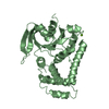
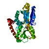
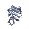
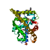
- PDB-1e3g: Human Androgen Receptor Ligand Binding in complex with the ligand... -
+ Open data

- Basic information
| Entry | Database: PDB / ID: 1e3g | |||||||||
|---|---|---|---|---|---|---|---|---|---|---|
| Title | Human Androgen Receptor Ligand Binding in complex with the ligand metribolone (R1881) | |||||||||
 Components Components | ANDROGEN RECEPTOR | |||||||||
 Keywords Keywords | ANDROGEN RECEPTOR / HUMAN ANDROGEN RECEPTOR / LIGAND BINDING DOMAIN | |||||||||
| Function / homology |  Function and homology information Function and homology informationPOU domain binding / positive regulation of integrin biosynthetic process / cellular response to testosterone stimulus / androgen binding / membraneless organelle assembly / RNA polymerase II general transcription initiation factor binding / androgen receptor signaling pathway / positive regulation of transcription by RNA polymerase III / cellular response to steroid hormone stimulus / nuclear steroid receptor activity ...POU domain binding / positive regulation of integrin biosynthetic process / cellular response to testosterone stimulus / androgen binding / membraneless organelle assembly / RNA polymerase II general transcription initiation factor binding / androgen receptor signaling pathway / positive regulation of transcription by RNA polymerase III / cellular response to steroid hormone stimulus / nuclear steroid receptor activity / RUNX2 regulates osteoblast differentiation / cellular response to estrogen stimulus / estrogen response element binding / RNA polymerase II core promoter sequence-specific DNA binding / regulation of protein localization to plasma membrane / estrogen receptor signaling pathway / intracellular receptor signaling pathway / steroid binding / HSP90 chaperone cycle for steroid hormone receptors (SHR) in the presence of ligand / negative regulation of extrinsic apoptotic signaling pathway / SUMOylation of intracellular receptors / RNA polymerase II transcription regulatory region sequence-specific DNA binding / positive regulation of cell differentiation / molecular condensate scaffold activity / Activated PKN1 stimulates transcription of AR (androgen receptor) regulated genes KLK2 and KLK3 / Nuclear Receptor transcription pathway / male gonad development / positive regulation of miRNA transcription / beta-catenin binding / nuclear receptor activity / transcription coactivator binding / cell-cell signaling / ATPase binding / DNA-binding transcription activator activity, RNA polymerase II-specific / spermatogenesis / molecular adaptor activity / RNA polymerase II-specific DNA-binding transcription factor binding / DNA-binding transcription factor activity, RNA polymerase II-specific / nuclear speck / transcription cis-regulatory region binding / Ub-specific processing proteases / RNA polymerase II cis-regulatory region sequence-specific DNA binding / DNA-binding transcription factor activity / negative regulation of cell population proliferation / signaling receptor binding / chromatin binding / positive regulation of cell population proliferation / positive regulation of gene expression / positive regulation of DNA-templated transcription / chromatin / enzyme binding / negative regulation of transcription by RNA polymerase II / signal transduction / DNA-templated transcription / positive regulation of transcription by RNA polymerase II / protein-containing complex / zinc ion binding / nucleoplasm / nucleus / plasma membrane / cytosol / cytoplasm Similarity search - Function | |||||||||
| Biological species |  HOMO SAPIENS (human) HOMO SAPIENS (human) | |||||||||
| Method |  X-RAY DIFFRACTION / SYNCHROTRON / MOLECULAR REPLACEMENT / Resolution: 2.4 Å X-RAY DIFFRACTION / SYNCHROTRON / MOLECULAR REPLACEMENT / Resolution: 2.4 Å | |||||||||
 Authors Authors | Matias, P.M. / Donner, P. / Coelho, R. / Thomaz, M. / Peixoto, C. / Macedo, S. / Otto, N. / Joschko, S. / Scholz, P. / Wegg, A. ...Matias, P.M. / Donner, P. / Coelho, R. / Thomaz, M. / Peixoto, C. / Macedo, S. / Otto, N. / Joschko, S. / Scholz, P. / Wegg, A. / Basler, S. / Schafer, M. / Ruff, M. / Egner, U. / Carrondo, M.A. | |||||||||
 Citation Citation | Journal: J. Biol. Chem. / Year: 2000 Title: Structural evidence for ligand specificity in the binding domain of the human androgen receptor. Implications for pathogenic gene mutations. Authors: Matias, P.M. / Donner, P. / Coelho, R. / Thomaz, M. / Peixoto, C. / Macedo, S. / Otto, N. / Joschko, S. / Scholz, P. / Wegg, A. / Basler, S. / Schafer, M. / Egner, U. / Carrondo, M.A. | |||||||||
| History |
|
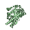
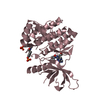
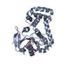
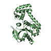
- Structure visualization
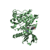
| Structure viewer | Molecule: MolmilJmol/JSmol |
|---|
- Downloads & links
-Download
| PDBx/mmCIF format | 1e3g.cif.gz | 66 KB | Display | PDBx/mmCIF format |
|---|---|---|---|---|
| PDB format | pdb1e3g.ent.gz | 47.4 KB | Display | PDB format |
| PDBx/mmJSON format | 1e3g.json.gz | Tree view | PDBx/mmJSON format | |
| Others |  Other downloads Other downloads |
-Validation report
| Arichive directory | https://data.pdbj.org/pub/pdb/validation_reports/e3/1e3gftp://data.pdbj.org/pub/pdb/validation_reports/e3/1e3g | HTTPS FTP |
|---|
-Related structure data
| Related structure data |  1e3kC 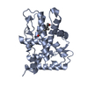 1a28S S: Starting model for refinement C: citing same article ( |
|---|---|
| Similar structure data |

-Links
 PDBj
PDBj


- Assembly
| Deposited unit | 
| ||||||||
|---|---|---|---|---|---|---|---|---|---|
| 1 |
| ||||||||
| Unit cell |
|
-Components
| #1: Protein | Mass: 30664.869 Da / Num. of mol.: 1 / Fragment: LIGAND-BINDING DOMAIN RESIDUES 447-709 Source method: isolated from a genetically manipulated source Source: (gene. exp.) HOMO SAPIENS (human) / Production host:  |
|---|---|
| #2: Chemical | ChemComp-R18 / (  Mass: 284.393 Da / Num. of mol.: 1 / Source method: obtained synthetically / Formula: C19H24O2 Mass: 284.393 Da / Num. of mol.: 1 / Source method: obtained synthetically / Formula: C19H24O2 |
| #3: Water | ChemComp-HOH /  Mass: 18.015 Da / Num. of mol.: 26 / Source method: isolated from a natural source / Formula: H2O Mass: 18.015 Da / Num. of mol.: 26 / Source method: isolated from a natural source / Formula: H2O |
| Has protein modification | Y |
-Experimental details
-Experiment
| Experiment | Method: X-RAY DIFFRACTION / Number of used crystals: 1 |
|---|
- Sample preparation
| Crystal | Density Matthews: 2.1 Å3/Da / Density % sol: 41.4 % | ||||||||||||||||||||||||||||||||||||||||||
|---|---|---|---|---|---|---|---|---|---|---|---|---|---|---|---|---|---|---|---|---|---|---|---|---|---|---|---|---|---|---|---|---|---|---|---|---|---|---|---|---|---|---|---|
| Crystal grow | Method: vapor diffusion, sitting drop / pH: 8.5 Details: RESERVOIR SOLUTION: 0.4M NA2HPO4-2(H2O), 0.4M K2HPO4, 0.1M TRIS-HCL PH 8.5, 0.1M (NH4)2HPO4 AND 5% PEG200. DROPS WERE COMPOSED OF EQUAL VOLUMES OF PROTEIN AND RESERVOIR SOLUTION AND WERE SET ...Details: RESERVOIR SOLUTION: 0.4M NA2HPO4-2(H2O), 0.4M K2HPO4, 0.1M TRIS-HCL PH 8.5, 0.1M (NH4)2HPO4 AND 5% PEG200. DROPS WERE COMPOSED OF EQUAL VOLUMES OF PROTEIN AND RESERVOIR SOLUTION AND WERE SET UP USING THE SITTING DROP METHOD. | ||||||||||||||||||||||||||||||||||||||||||
| Crystal grow | *PLUS Temperature: 20 ℃ / Method: vapor diffusion, sitting drop | ||||||||||||||||||||||||||||||||||||||||||
| Components of the solutions | *PLUS
|
-Data collection
| Diffraction | Mean temperature: 100 K |
|---|---|
| Diffraction source | Source: SYNCHROTRON / Site: ESRF  / Beamline: ID14-4 / Wavelength: 0.932 / Beamline: ID14-4 / Wavelength: 0.932 |
| Detector | Type: ADSC CCD / Detector: CCD / Date: Sep 15, 1999 / Details: TOROIDAL MIRRORS |
| Radiation | Monochromator: DOUBLE CRYSTAL SI 111 / Protocol: SINGLE WAVELENGTH / Monochromatic (M) / Laue (L): M / Scattering type: x-ray |
| Radiation wavelength | Wavelength: 0.932 Å / Relative weight: 1 |
| Reflection | Resolution: 2.4→24.4 Å / Num. obs: 10638 / % possible obs: 99.8 % / Redundancy: 3.5 % / Biso Wilson estimate: 49.4 Å2 / Rmerge(I) obs: 0.078 |
| Reflection shell | Resolution: 2.4→2.46 Å / Rmerge(I) obs: 0.351 / % possible all: 99.9 |
| Reflection | *PLUS Num. measured all: 37443 |
| Reflection shell | *PLUS % possible obs: 99.9 % |
- Processing
| Software |
| ||||||||||||||||||||||||||||||||||||||||||||||||||||||||||||
|---|---|---|---|---|---|---|---|---|---|---|---|---|---|---|---|---|---|---|---|---|---|---|---|---|---|---|---|---|---|---|---|---|---|---|---|---|---|---|---|---|---|---|---|---|---|---|---|---|---|---|---|---|---|---|---|---|---|---|---|---|---|
| Refinement | Method to determine structure: MOLECULAR REPLACEMENT Starting model: PDB ENTRY 1A28 Resolution: 2.4→24.4 Å / Isotropic thermal model: RESTRAINED / Cross valid method: THROUGHOUT / σ(F): 2 Details: C-TERMINAL RESIDUE WAS NOT SEEN IN THE DENSITY MAPS
| ||||||||||||||||||||||||||||||||||||||||||||||||||||||||||||
| Displacement parameters | Biso mean: 45.8 Å2 | ||||||||||||||||||||||||||||||||||||||||||||||||||||||||||||
| Refine analyze | Luzzati d res low obs: 25 Å / Luzzati sigma a obs: 0.47 Å | ||||||||||||||||||||||||||||||||||||||||||||||||||||||||||||
| Refinement step | Cycle: LAST / Resolution: 2.4→24.4 Å
| ||||||||||||||||||||||||||||||||||||||||||||||||||||||||||||
| Refine LS restraints |
| ||||||||||||||||||||||||||||||||||||||||||||||||||||||||||||
| Xplor file |
| ||||||||||||||||||||||||||||||||||||||||||||||||||||||||||||
| Software | *PLUS Name: X-PLOR / Version: 3.1 / Classification: refinement | ||||||||||||||||||||||||||||||||||||||||||||||||||||||||||||
| Refinement | *PLUS Rfactor obs: 0.21 / Rfactor Rwork: 0.21 | ||||||||||||||||||||||||||||||||||||||||||||||||||||||||||||
| Solvent computation | *PLUS | ||||||||||||||||||||||||||||||||||||||||||||||||||||||||||||
| Displacement parameters | *PLUS | ||||||||||||||||||||||||||||||||||||||||||||||||||||||||||||
| Refine LS restraints | *PLUS
|
