 Movie
Movie Controller

[English] 日本語
 Yorodumi
- PDB-1e3k: Human Progesteron Receptor Ligand Binding Domain in complex with ... -
+ Open data

- Basic information
| Entry | Database: PDB / ID: 1e3k | ||||||
|---|---|---|---|---|---|---|---|
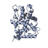
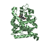
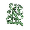
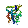
| Title | Human Progesteron Receptor Ligand Binding Domain in complex with the ligand metribolone (R1881) | ||||||
 Components Components | PROGESTERONE RECEPTOR | ||||||
 Keywords Keywords | HUMAN PROGESTERONE RECEPTOR / LIGAND BINDING DOMAIN | ||||||
| Function / homology |  Function and homology information Function and homology informationglandular epithelial cell maturation / tertiary branching involved in mammary gland duct morphogenesis / ovulation from ovarian follicle / paracrine signaling / regulation of epithelial cell proliferation / maintenance of protein location in nucleus / lung alveolus development / nuclear steroid receptor activity / progesterone receptor signaling pathway / nuclear receptor-mediated steroid hormone signaling pathway ...glandular epithelial cell maturation / tertiary branching involved in mammary gland duct morphogenesis / ovulation from ovarian follicle / paracrine signaling / regulation of epithelial cell proliferation / maintenance of protein location in nucleus / lung alveolus development / nuclear steroid receptor activity / progesterone receptor signaling pathway / nuclear receptor-mediated steroid hormone signaling pathway / estrogen response element binding / Nuclear signaling by ERBB4 / steroid binding / HSP90 chaperone cycle for steroid hormone receptors (SHR) in the presence of ligand / SUMOylation of intracellular receptors / Nuclear Receptor transcription pathway / nuclear receptor activity / transcription coactivator binding / cell-cell signaling / ATPase binding / DNA-binding transcription activator activity, RNA polymerase II-specific / Estrogen-dependent gene expression / DNA-binding transcription factor activity, RNA polymerase II-specific / mitochondrial outer membrane / RNA polymerase II cis-regulatory region sequence-specific DNA binding / signaling receptor binding / negative regulation of gene expression / positive regulation of gene expression / regulation of transcription by RNA polymerase II / regulation of DNA-templated transcription / chromatin / enzyme binding / signal transduction / positive regulation of transcription by RNA polymerase II / DNA binding / zinc ion binding / nucleoplasm / identical protein binding / nucleus / plasma membrane / cytosol Similarity search - Function | ||||||
| Biological species |  HOMO SAPIENS (human) HOMO SAPIENS (human) | ||||||
| Method |  X-RAY DIFFRACTION / SYNCHROTRON / MOLECULAR REPLACEMENT / Resolution: 2.8 Å X-RAY DIFFRACTION / SYNCHROTRON / MOLECULAR REPLACEMENT / Resolution: 2.8 Å | ||||||
 Authors Authors | Matias, P.M. / Donner, P. / Coelho, R. / Thomaz, M. / Peixoto, C. / Macedo, S. / Otto, N. / Joschko, S. / Scholz, P. / Wegg, A. ...Matias, P.M. / Donner, P. / Coelho, R. / Thomaz, M. / Peixoto, C. / Macedo, S. / Otto, N. / Joschko, S. / Scholz, P. / Wegg, A. / Basler, S. / Schafer, M. / Egner, U. / Carrondo, M.A. | ||||||
 Citation Citation | Journal: J. Biol. Chem. / Year: 2000 Title: Structural evidence for ligand specificity in the binding domain of the human androgen receptor. Implications for pathogenic gene mutations. Authors: Matias, P.M. / Donner, P. / Coelho, R. / Thomaz, M. / Peixoto, C. / Macedo, S. / Otto, N. / Joschko, S. / Scholz, P. / Wegg, A. / Basler, S. / Schafer, M. / Egner, U. / Carrondo, M.A. | ||||||
| History |
|
- Structure visualization
| Structure viewer | Molecule: MolmilJmol/JSmol |
|---|
- Downloads & links
-Download
| PDBx/mmCIF format | 1e3k.cif.gz | 118.1 KB | Display | PDBx/mmCIF format |
|---|---|---|---|---|
| PDB format | pdb1e3k.ent.gz | 87.4 KB | Display | PDB format |
| PDBx/mmJSON format | 1e3k.json.gz | Tree view | PDBx/mmJSON format | |
| Others |  Other downloads Other downloads |
-Validation report
| Arichive directory | https://data.pdbj.org/pub/pdb/validation_reports/e3/1e3kftp://data.pdbj.org/pub/pdb/validation_reports/e3/1e3k | HTTPS FTP |
|---|
-Related structure data
| Related structure data |  1e3gC  1a28S S: Starting model for refinement C: citing same article ( |
|---|---|
| Similar structure data |


-Links
 PDBj
PDBj


- Assembly
| Deposited unit | 
| ||||||||
|---|---|---|---|---|---|---|---|---|---|
| 1 | 
| ||||||||
| 2 | 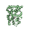
| ||||||||
| Unit cell |
| ||||||||
| Noncrystallographic symmetry (NCS) | NCS oper: (Code: given Matrix: (0.52983, 0.82973, 0.17561), Vector: |
-Components
| #1: Protein | Mass: 29738.824 Da / Num. of mol.: 2 / Fragment: LIGAND-BINDING DOMAIN Source method: isolated from a genetically manipulated source Source: (gene. exp.) HOMO SAPIENS (human) / Production host:  #2: Chemical |   Mass: 284.393 Da / Num. of mol.: 2 / Source method: obtained synthetically / Formula: C19H24O2 Mass: 284.393 Da / Num. of mol.: 2 / Source method: obtained synthetically / Formula: C19H24O2#3: Water | ChemComp-HOH / |  Mass: 18.015 Da / Num. of mol.: 1 / Source method: isolated from a natural source / Formula: H2O Mass: 18.015 Da / Num. of mol.: 1 / Source method: isolated from a natural source / Formula: H2O |
|---|
-Experimental details
-Experiment
| Experiment | Method: X-RAY DIFFRACTION / Number of used crystals: 1 |
|---|
- Sample preparation
| Crystal | Density Matthews: 2.26 Å3/Da / Density % sol: 45.6 % | |||||||||||||||||||||||||
|---|---|---|---|---|---|---|---|---|---|---|---|---|---|---|---|---|---|---|---|---|---|---|---|---|---|---|
| Crystal grow | Method: vapor diffusion, hanging drop / pH: 7.5 Details: RESERVOIR SOLUTION: 10% ISO-PROPANOL, 100MM SODIUM CITRATE 50MM HEPES PH 7.5. THE DROPS WERE SET UP USING THE HANGING DROP METHOD AND WERE COMPOSED OF A 2:1 RATIO OF PROTEIN AND RESERVOIR SOLUTIONS. | |||||||||||||||||||||||||
| Crystal grow | *PLUS Temperature: 4 ℃ / Method: vapor diffusion, hanging drop | |||||||||||||||||||||||||
| Components of the solutions | *PLUS
|
-Data collection
| Diffraction | Mean temperature: 100 K |
|---|---|
| Diffraction source | Source: SYNCHROTRON / Site: ESRF  / Beamline: ID14-4 / Wavelength: 0.9537 / Beamline: ID14-4 / Wavelength: 0.9537 |
| Detector | Type: MARRESEARCH / Detector: CCD / Details: MIRRORS |
| Radiation | Monochromator: DOUBLE CRYSTAL SI 111 / Protocol: SINGLE WAVELENGTH / Monochromatic (M) / Laue (L): M / Scattering type: x-ray |
| Radiation wavelength | Wavelength: 0.9537 Å / Relative weight: 1 |
| Reflection | Resolution: 2.8→12.5 Å / Num. obs: 8875 / % possible obs: 67 % / Redundancy: 7.6 % / Biso Wilson estimate: 48.2 Å2 / Rmerge(I) obs: 0.048 / Net I/σ(I): 15.2 |
| Reflection shell | Resolution: 2.8→2.87 Å / Rmerge(I) obs: 0.151 / % possible all: 68.8 |
| Reflection | *PLUS Num. measured all: 67655 |
| Reflection shell | *PLUS % possible obs: 68.8 % |
- Processing
| Software |
| |||||||||||||||||||||||||||||||||||||||||||||||||||||||||||||||
|---|---|---|---|---|---|---|---|---|---|---|---|---|---|---|---|---|---|---|---|---|---|---|---|---|---|---|---|---|---|---|---|---|---|---|---|---|---|---|---|---|---|---|---|---|---|---|---|---|---|---|---|---|---|---|---|---|---|---|---|---|---|---|---|---|
| Refinement | Method to determine structure: MOLECULAR REPLACEMENT Starting model: PDB ENTRY 1A28 Resolution: 2.8→12.5 Å / Cross valid method: THROUGHOUT / σ(F): 0 / Details: FINAL 2 C-TERMINAL RESIDUES NOT SEEN IN THE MAP
| |||||||||||||||||||||||||||||||||||||||||||||||||||||||||||||||
| Refinement step | Cycle: LAST / Resolution: 2.8→12.5 Å
| |||||||||||||||||||||||||||||||||||||||||||||||||||||||||||||||
| Refine LS restraints |
| |||||||||||||||||||||||||||||||||||||||||||||||||||||||||||||||
| Software | *PLUS Name: REFMAC / Classification: refinement | |||||||||||||||||||||||||||||||||||||||||||||||||||||||||||||||
| Refinement | *PLUS Rfactor obs: 0.217 | |||||||||||||||||||||||||||||||||||||||||||||||||||||||||||||||
| Solvent computation | *PLUS | |||||||||||||||||||||||||||||||||||||||||||||||||||||||||||||||
| Displacement parameters | *PLUS |
