 Movie
Movie Controller

[English] 日本語
 Yorodumi
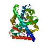
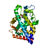
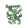
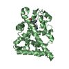
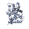
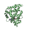
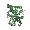
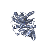
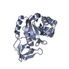
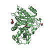
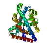
- PDB-1i37: CRYSTAL STRUCTURE OF THE RAT ANDROGEN RECEPTOR LIGAND BINDING DOM... -
+ Open data

- Basic information
| Entry | Database: PDB / ID: 1i37 | ||||||
|---|---|---|---|---|---|---|---|
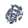
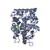
| Title | CRYSTAL STRUCTURE OF THE RAT ANDROGEN RECEPTOR LIGAND BINDING DOMAIN COMPLEX WITH DIHYDROTESTOSTERONE | ||||||
 Components Components | ANDROGEN RECEPTOR | ||||||
 Keywords Keywords | HORMONE/GROWTH FACTOR / ANDROGEN RECEPTOR / STEROID RECEPTOR / NUCLEAR RECEPTOR / TRANSCRIPTION REGULATION / LIGAND-BINDING DOMAIN / HORMONE-GROWTH FACTOR COMPLEX | ||||||
| Function / homology |  Function and homology information Function and homology informationActivated PKN1 stimulates transcription of AR (androgen receptor) regulated genes KLK2 and KLK3 / copulation / male sex differentiation / skeletal muscle hypertrophy / HSP90 chaperone cycle for steroid hormone receptors (SHR) in the presence of ligand / regulation of prostatic bud formation / positive regulation of penile erection / ribonucleotide binding / male courtship behavior / positive regulation of hippocampal neuron apoptotic process ...Activated PKN1 stimulates transcription of AR (androgen receptor) regulated genes KLK2 and KLK3 / copulation / male sex differentiation / skeletal muscle hypertrophy / HSP90 chaperone cycle for steroid hormone receptors (SHR) in the presence of ligand / regulation of prostatic bud formation / positive regulation of penile erection / ribonucleotide binding / male courtship behavior / positive regulation of hippocampal neuron apoptotic process / male somatic sex determination / prostate induction / Ub-specific processing proteases / lateral sprouting involved in mammary gland duct morphogenesis / regulation of developmental growth / POU domain binding / male genitalia morphogenesis / positive regulation of integrin biosynthetic process / reproductive structure development / tertiary branching involved in mammary gland duct morphogenesis / SUMOylation of intracellular receptors / Nuclear Receptor transcription pathway / cellular response to testosterone stimulus / animal organ formation / positive regulation of locomotion / androgen binding / cellular response to follicle-stimulating hormone stimulus / regulation of systemic arterial blood pressure / response to environmental enrichment / Leydig cell differentiation / reproductive behavior / epithelial cell differentiation involved in prostate gland development / positive regulation of epithelial cell proliferation involved in prostate gland development / drinking behavior / prostate gland epithelium morphogenesis / prostate gland growth / epithelial cell morphogenesis / membraneless organelle assembly / reproductive system development / RNA polymerase II general transcription initiation factor binding / positive regulation of insulin-like growth factor receptor signaling pathway / fertilization / response to steroid hormone / locomotion / androgen receptor signaling pathway / positive regulation of transcription by RNA polymerase III / nuclear androgen receptor binding / morphogenesis of an epithelial fold / cellular response to steroid hormone stimulus / positive regulation of intracellular estrogen receptor signaling pathway / response to muscle activity / response to testosterone / seminiferous tubule development / nuclear steroid receptor activity / mammary gland alveolus development / cellular response to estrogen stimulus / estrogen response element binding / single fertilization / RNA polymerase II core promoter sequence-specific DNA binding / regulation of protein localization to plasma membrane / estrogen receptor signaling pathway / intracellular receptor signaling pathway / steroid binding / liver regeneration / negative regulation of extrinsic apoptotic signaling pathway / response to nicotine / RNA polymerase II transcription regulatory region sequence-specific DNA binding / positive regulation of cell differentiation / molecular condensate scaffold activity / response to insulin / positive regulation of miRNA transcription / beta-catenin binding / male gonad development / nuclear receptor activity / multicellular organism growth / transcription coactivator binding / negative regulation of epithelial cell proliferation / nuclear matrix / response to estradiol / ATPase binding / regulation of gene expression / DNA-binding transcription activator activity, RNA polymerase II-specific / spermatogenesis / sequence-specific DNA binding / gene expression / molecular adaptor activity / in utero embryonic development / RNA polymerase II-specific DNA-binding transcription factor binding / response to ethanol / positive regulation of MAPK cascade / transcription cis-regulatory region binding / nuclear speck / RNA polymerase II cis-regulatory region sequence-specific DNA binding / DNA-binding transcription factor activity / signaling receptor binding / negative regulation of cell population proliferation / protein domain specific binding / axon / negative regulation of DNA-templated transcription / positive regulation of cell population proliferation Similarity search - Function | ||||||
| Biological species |  | ||||||
| Method |  X-RAY DIFFRACTION / SYNCHROTRON / MOLECULAR REPLACEMENT / Resolution: 2 Å X-RAY DIFFRACTION / SYNCHROTRON / MOLECULAR REPLACEMENT / Resolution: 2 Å | ||||||
 Authors Authors | Sack, J.S. | ||||||
 Citation Citation | Journal: Proc.Natl.Acad.Sci.USA / Year: 2001 Title: Crystallographic structures of the ligand-binding domains of the androgen receptor and its T877A mutant complexed with the natural agonist dihydrotestosterone. Authors: Sack, J.S. / Kish, K.F. / Wang, C. / Attar, R.M. / Kiefer, S.E. / An, Y. / Wu, G.Y. / Scheffler, J.E. / Salvati, M.E. / Krystek Jr., S.R. / Weinmann, R. / Einspahr, H.M. | ||||||
| History |
|
- Structure visualization
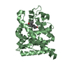
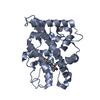
| Structure viewer | Molecule: MolmilJmol/JSmol |
|---|
- Downloads & links
-Download
| PDBx/mmCIF format | 1i37.cif.gz | 66 KB | Display | PDBx/mmCIF format |
|---|---|---|---|---|
| PDB format | pdb1i37.ent.gz | 47.2 KB | Display | PDB format |
| PDBx/mmJSON format | 1i37.json.gz | Tree view | PDBx/mmJSON format | |
| Others |  Other downloads Other downloads |
-Validation report
| Arichive directory | https://data.pdbj.org/pub/pdb/validation_reports/i3/1i37ftp://data.pdbj.org/pub/pdb/validation_reports/i3/1i37 | HTTPS FTP |
|---|
-Related structure data
| Related structure data | 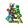 1i38C  1a28S C: citing same article ( S: Starting model for refinement |
|---|---|
| Similar structure data |

-Links
 PDBj
PDBj

- Assembly
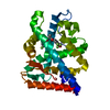
| Deposited unit | 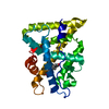
| ||||||||
|---|---|---|---|---|---|---|---|---|---|
| 1 |
| ||||||||
| Unit cell |
|
-Components
| #1: Protein | Mass: 30282.414 Da / Num. of mol.: 1 / Fragment: LIGAND-BINDING DOMAIN Source method: isolated from a genetically manipulated source Source: (gene. exp.)  |
|---|---|
| #2: Chemical | ChemComp-DHT / 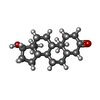  Mass: 290.440 Da / Num. of mol.: 1 / Source method: obtained synthetically / Formula: C19H30O2 / Comment: hormone*YM Mass: 290.440 Da / Num. of mol.: 1 / Source method: obtained synthetically / Formula: C19H30O2 / Comment: hormone*YM |
| #3: Water | ChemComp-HOH /  Mass: 18.015 Da / Num. of mol.: 106 / Source method: isolated from a natural source / Formula: H2O Mass: 18.015 Da / Num. of mol.: 106 / Source method: isolated from a natural source / Formula: H2O |
-Experimental details
-Experiment
| Experiment | Method: X-RAY DIFFRACTION / Number of used crystals: 1 |
|---|
- Sample preparation
| Crystal | Density Matthews: 2.33 Å3/Da / Density % sol: 50.2 % | ||||||||||||||||||||
|---|---|---|---|---|---|---|---|---|---|---|---|---|---|---|---|---|---|---|---|---|---|
| Crystal grow | Temperature: 297 K / Method: vapor diffusion, hanging drop / pH: 7.5 Details: 0.88 M NA TARTRATE, 0.1M NA HEPES, pH 7.5, VAPOR DIFFUSION, HANGING DROP, temperature 297K | ||||||||||||||||||||
| Crystal grow | *PLUS Temperature: 20 ℃Details: drop consists of equal amounts of protein and reservoir solutions | ||||||||||||||||||||
| Components of the solutions | *PLUS
|
-Data collection
| Diffraction | Mean temperature: 100 K |
|---|---|
| Diffraction source | Source: SYNCHROTRON / Site: APS  / Beamline: 17-ID / Wavelength: 1 / Beamline: 17-ID / Wavelength: 1 |
| Detector | Type: BRUKER / Detector: CCD / Date: Jun 17, 1999 |
| Radiation | Protocol: SINGLE WAVELENGTH / Monochromatic (M) / Laue (L): M / Scattering type: x-ray |
| Radiation wavelength | Wavelength: 1 Å / Relative weight: 1 |
| Reflection | Resolution: 2→50 Å / Num. obs: 17329 / % possible obs: 94.8 % / Redundancy: 7.3 % / Biso Wilson estimate: 29.3 Å2 / Rsym value: 0.101 / Net I/σ(I): 12 |
| Reflection shell | Resolution: 2→2.1 Å / Redundancy: 5.5 % / Mean I/σ(I) obs: 4 / Rsym value: 0.256 / % possible all: 73 |
| Reflection | *PLUS Rmerge(I) obs: 0.101 |
| Reflection shell | *PLUS % possible obs: 73 % / Rmerge(I) obs: 0.256 |
- Processing
| Software |
| ||||||||||||||||||||||||||||||||||||||||||||||||||||||||||||
|---|---|---|---|---|---|---|---|---|---|---|---|---|---|---|---|---|---|---|---|---|---|---|---|---|---|---|---|---|---|---|---|---|---|---|---|---|---|---|---|---|---|---|---|---|---|---|---|---|---|---|---|---|---|---|---|---|---|---|---|---|---|
| Refinement | Method to determine structure: MOLECULAR REPLACEMENT Starting model: PDB ENTRY 1A28 Resolution: 2→10 Å / Data cutoff high absF: 999999 / Data cutoff low absF: 0 / Cross valid method: THROUGHOUT / σ(F): 2 Details: The side chains of residues 845-850 were not seen in the density. HIS-TAG (RESIDUES 664 TO 671) AND RESIDUES 918 TO 919 WERE NOT SEEN IN THE ELECTRON DENSITY AND ARE PRESUMED TO BE DISORDERED.
| ||||||||||||||||||||||||||||||||||||||||||||||||||||||||||||
| Displacement parameters | Biso mean: 25.2 Å2 | ||||||||||||||||||||||||||||||||||||||||||||||||||||||||||||
| Refine analyze | Luzzati coordinate error obs: 0.34 Å | ||||||||||||||||||||||||||||||||||||||||||||||||||||||||||||
| Refinement step | Cycle: LAST / Resolution: 2→10 Å
| ||||||||||||||||||||||||||||||||||||||||||||||||||||||||||||
| Refine LS restraints |
| ||||||||||||||||||||||||||||||||||||||||||||||||||||||||||||
| LS refinement shell | Resolution: 2→2.09 Å / Total num. of bins used: 8
| ||||||||||||||||||||||||||||||||||||||||||||||||||||||||||||
| Software | *PLUS Name: X-PLOR / Classification: refinement | ||||||||||||||||||||||||||||||||||||||||||||||||||||||||||||
| Refine LS restraints | *PLUS
|
