[English] 日本語
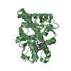
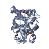
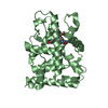
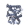
Yorodumi- PDB-4apu: PR X-Ray structures in agonist conformations reveal two different... -
+Open data
-Basic information
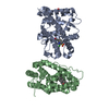
Entry | Database: PDB / ID: 4apu | ||||||
---|---|---|---|---|---|---|---|
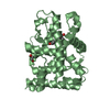
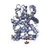
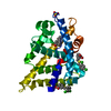
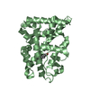
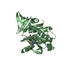
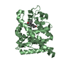
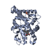
Title | PR X-Ray structures in agonist conformations reveal two different mechanisms for partial agonism in 11beta-substituted steroids | ||||||
Components | PROGESTERONE RECEPTOR | ||||||
Keywords | TRANSCRIPTION / ASOPRISNIL / PARTIAL AGONIST | ||||||
Function / homology | Function and homology information glandular epithelial cell maturation / tertiary branching involved in mammary gland duct morphogenesis / ovulation from ovarian follicle / paracrine signaling / maintenance of protein location in nucleus / regulation of epithelial cell proliferation / nuclear steroid receptor activity / lung alveolus development / estrogen response element binding / progesterone receptor signaling pathway ...glandular epithelial cell maturation / tertiary branching involved in mammary gland duct morphogenesis / ovulation from ovarian follicle / paracrine signaling / maintenance of protein location in nucleus / regulation of epithelial cell proliferation / nuclear steroid receptor activity / lung alveolus development / estrogen response element binding / progesterone receptor signaling pathway / nuclear receptor-mediated steroid hormone signaling pathway / Nuclear signaling by ERBB4 / HSP90 chaperone cycle for steroid hormone receptors (SHR) in the presence of ligand / steroid binding / G protein-coupled receptor activity / SUMOylation of intracellular receptors / transcription coactivator binding / Nuclear Receptor transcription pathway / nuclear receptor activity / cell-cell signaling / ATPase binding / DNA-binding transcription activator activity, RNA polymerase II-specific / Estrogen-dependent gene expression / mitochondrial outer membrane / DNA-binding transcription factor activity, RNA polymerase II-specific / RNA polymerase II cis-regulatory region sequence-specific DNA binding / negative regulation of gene expression / signaling receptor binding / regulation of DNA-templated transcription / positive regulation of gene expression / chromatin / regulation of transcription by RNA polymerase II / enzyme binding / signal transduction / positive regulation of transcription by RNA polymerase II / DNA binding / zinc ion binding / nucleoplasm / identical protein binding / plasma membrane / cytosol Similarity search - Function | ||||||
Biological species | HOMO SAPIENS (human) | ||||||
Method | X-RAY DIFFRACTION / FOURIER SYNTHESIS / Resolution: 1.9 Å | ||||||
Authors | Lusher, S.J. / Raaijmakers, H.C.A. / Bosch, R. / Vu-Pham, D. / McGuire, R. / Oubrie, A. / de Vlieg, J. | ||||||
Citation | Journal: J. Biol. Chem. / Year: 2012 Title: X-ray structures of progesterone receptor ligand binding domain in its agonist state reveal differing mechanisms for mixed profiles of 11 beta-substituted steroids. Authors: Lusher, S.J. / Raaijmakers, H.C. / Vu-Pham, D. / Kazemier, B. / Bosch, R. / McGuire, R. / Azevedo, R. / Hamersma, H. / Dechering, K. / Oubrie, A. / van Duin, M. / de Vlieg, J. | ||||||
History |
|
-Structure visualization
Structure viewer | Molecule: MolmilJmol/JSmol |
---|
-Downloads & links
-Download
PDBx/mmCIF format | 4apu.cif.gz | 223.6 KB | Display | PDBx/mmCIF format |
---|---|---|---|---|
PDB format | pdb4apu.ent.gz | 179 KB | Display | PDB format |
PDBx/mmJSON format | 4apu.json.gz | Tree view | PDBx/mmJSON format | |
Others | Other downloads |
-Validation report
Summary document | 4apu_validation.pdf.gz | 1.2 MB | Display | wwPDB validaton report |
---|---|---|---|---|
Full document | 4apu_full_validation.pdf.gz | 1.2 MB | Display | |
Data in XML | 4apu_validation.xml.gz | 21.8 KB | Display | |
Data in CIF | 4apu_validation.cif.gz | 30.5 KB | Display | |
Arichive directory | https://data.pdbj.org/pub/pdb/validation_reports/ap/4apu ftp://data.pdbj.org/pub/pdb/validation_reports/ap/4apu | HTTPS FTP |
-Related structure data
Related structure data | 4a2jC 3zr7S S: Starting model for refinement C: citing same article (ref.) |
---|---|
Similar structure data |
-Links
-Assembly
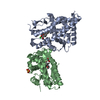
Deposited unit |
| ||||||||
---|---|---|---|---|---|---|---|---|---|
1 |
| ||||||||
2 |
| ||||||||
Unit cell |
|
-Components
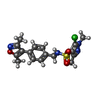
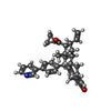
#1: Protein | Mass: 29968.098 Da / Num. of mol.: 2 / Fragment: LIGAND BINDING DOMAIN, RESIDUES 514-769 Source method: isolated from a genetically manipulated source Source: (gene. exp.) HOMO SAPIENS (human) / Production host: ESCHERICHIA COLI (E. coli) / Strain (production host): BL21(DE3) / References: UniProt: P06401 #2: Chemical | ChemComp-OR8 / | #3: Chemical | ChemComp-SO4 / | #4: Chemical | ChemComp-A2K / ( | #5: Water | ChemComp-HOH / | |
---|
-Experimental details
-Experiment
Experiment | Method: X-RAY DIFFRACTION / Number of used crystals: 1 |
---|
-Sample preparation
Crystal | Density Matthews: 2.31 Å3/Da / Density % sol: 46.8 % |
---|---|
Crystal grow | pH: 6.5 Details: 20-30% POLYETHYLENE GLYCOL 3350, 0.1 M HEPES PH 6.5, 100 MM MG2SO4, 10% GLYCEROL |
-Data collection
Diffraction | Mean temperature: 100 K |
---|---|
Diffraction source | Source: ROTATING ANODE / Type: RIGAKU / Wavelength: 1.5418 |
Detector | Type: R-AXIS / Detector: IMAGE PLATE / Date: Mar 10, 2011 / Details: MIRRORS |
Radiation | Protocol: SINGLE WAVELENGTH / Monochromatic (M) / Laue (L): M / Scattering type: x-ray |
Radiation wavelength | Wavelength: 1.5418 Å / Relative weight: 1 |
Reflection | Resolution: 1.9→47.4 Å / Num. obs: 40585 / % possible obs: 100 % / Observed criterion σ(I): 0 / Redundancy: 4.1 % / Rmerge(I) obs: 0.08 / Net I/σ(I): 9.3 |
Reflection shell | Resolution: 1.9→2 Å / Redundancy: 4 % / Rmerge(I) obs: 0.73 / Mean I/σ(I) obs: 1.9 / % possible all: 99.9 |
-Processing
Software |
| ||||||||||||||||||||||||||||||||||||||||||||||||||||||||||||||||||||||||||||||||||||||||||||||||||||||||||||||||||||||||||||||||||||||||||||||||||||||||||||||||||||||||||||||||||||||
---|---|---|---|---|---|---|---|---|---|---|---|---|---|---|---|---|---|---|---|---|---|---|---|---|---|---|---|---|---|---|---|---|---|---|---|---|---|---|---|---|---|---|---|---|---|---|---|---|---|---|---|---|---|---|---|---|---|---|---|---|---|---|---|---|---|---|---|---|---|---|---|---|---|---|---|---|---|---|---|---|---|---|---|---|---|---|---|---|---|---|---|---|---|---|---|---|---|---|---|---|---|---|---|---|---|---|---|---|---|---|---|---|---|---|---|---|---|---|---|---|---|---|---|---|---|---|---|---|---|---|---|---|---|---|---|---|---|---|---|---|---|---|---|---|---|---|---|---|---|---|---|---|---|---|---|---|---|---|---|---|---|---|---|---|---|---|---|---|---|---|---|---|---|---|---|---|---|---|---|---|---|---|---|
Refinement | Method to determine structure: FOURIER SYNTHESIS Starting model: PDB ENTRY 3ZR7 Resolution: 1.9→70.04 Å / Cor.coef. Fo:Fc: 0.965 / Cor.coef. Fo:Fc free: 0.945 / SU B: 12.436 / SU ML: 0.151 / Cross valid method: THROUGHOUT / ESU R: 0.178 / ESU R Free: 0.162 / Stereochemistry target values: MAXIMUM LIKELIHOOD / Details: HYDROGENS HAVE BEEN ADDED IN THE RIDING POSITIONS.
| ||||||||||||||||||||||||||||||||||||||||||||||||||||||||||||||||||||||||||||||||||||||||||||||||||||||||||||||||||||||||||||||||||||||||||||||||||||||||||||||||||||||||||||||||||||||
Solvent computation | Ion probe radii: 0.8 Å / Shrinkage radii: 0.8 Å / VDW probe radii: 1.2 Å / Solvent model: MASK | ||||||||||||||||||||||||||||||||||||||||||||||||||||||||||||||||||||||||||||||||||||||||||||||||||||||||||||||||||||||||||||||||||||||||||||||||||||||||||||||||||||||||||||||||||||||
Displacement parameters | Biso mean: 35.637 Å2
| ||||||||||||||||||||||||||||||||||||||||||||||||||||||||||||||||||||||||||||||||||||||||||||||||||||||||||||||||||||||||||||||||||||||||||||||||||||||||||||||||||||||||||||||||||||||
Refinement step | Cycle: LAST / Resolution: 1.9→70.04 Å
| ||||||||||||||||||||||||||||||||||||||||||||||||||||||||||||||||||||||||||||||||||||||||||||||||||||||||||||||||||||||||||||||||||||||||||||||||||||||||||||||||||||||||||||||||||||||
Refine LS restraints |
|