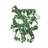
[English] 日本語
Yorodumi
- PDB-3zra: Structural basis for agonism and antagonism for a set of chemical... -

+
Open data


ID or keywords:

Loading...

-
Basic information

Entry
Database: PDB / ID: 3zra
TitleStructural basis for agonism and antagonism for a set of chemically related progesterone receptor modulators
ComponentsPROGESTERONE RECEPTOR
KeywordsHORMONE RECEPTOR
Function / homology
Function and homology information


glandular epithelial cell maturation / tertiary branching involved in mammary gland duct morphogenesis / ovulation from ovarian follicle / paracrine signaling / regulation of epithelial cell proliferation / nuclear steroid receptor activity / lung alveolus development / progesterone receptor signaling pathway / estrogen response element binding / Nuclear signaling by ERBB4 ...glandular epithelial cell maturation / tertiary branching involved in mammary gland duct morphogenesis / ovulation from ovarian follicle / paracrine signaling / regulation of epithelial cell proliferation / nuclear steroid receptor activity / lung alveolus development / progesterone receptor signaling pathway / estrogen response element binding / Nuclear signaling by ERBB4 / intracellular steroid hormone receptor signaling pathway / HSP90 chaperone cycle for steroid hormone receptors (SHR) in the presence of ligand / steroid binding / G protein-coupled receptor activity / SUMOylation of intracellular receptors / transcription coactivator binding / Nuclear Receptor transcription pathway / nuclear receptor activity / cell-cell signaling / ATPase binding / DNA-binding transcription activator activity, RNA polymerase II-specific / Estrogen-dependent gene expression / mitochondrial outer membrane / nucleic acid binding / DNA-binding transcription factor activity, RNA polymerase II-specific / RNA polymerase II cis-regulatory region sequence-specific DNA binding / negative regulation of gene expression / signaling receptor binding / chromatin / positive regulation of gene expression / regulation of transcription by RNA polymerase II / enzyme binding / signal transduction / positive regulation of transcription by RNA polymerase II / DNA binding / zinc ion binding / nucleoplasm / identical protein binding / plasma membrane / cytosol
Similarity search - Function
Progesterone receptor / Progesterone receptor / Retinoid X Receptor / Retinoid X Receptor / Nuclear hormone receptor / Nuclear hormones receptors DNA-binding region signature. / Zinc finger, nuclear hormone receptor-type / Zinc finger, C4 type (two domains) / Nuclear hormone receptors DNA-binding domain profile. / c4 zinc finger in nuclear hormone receptors ...Progesterone receptor / Progesterone receptor / Retinoid X Receptor / Retinoid X Receptor / Nuclear hormone receptor / Nuclear hormones receptors DNA-binding region signature. / Zinc finger, nuclear hormone receptor-type / Zinc finger, C4 type (two domains) / Nuclear hormone receptors DNA-binding domain profile. / c4 zinc finger in nuclear hormone receptors / Nuclear hormone receptor, ligand-binding domain / Nuclear hormone receptor-like domain superfamily / Ligand-binding domain of nuclear hormone receptor / Nuclear receptor (NR) ligand-binding (LBD) domain profile. / Ligand binding domain of hormone receptors / Zinc finger, NHR/GATA-type / Orthogonal Bundle / Mainly Alpha
Similarity search - Domain/homology
Chem-ORB / Progesterone receptor
Similarity search - Component
Biological speciesHOMO SAPIENS (human)
MethodX-RAY DIFFRACTION / SYNCHROTRON / OTHER / Resolution: 1.9 Å
AuthorsLusher, S.J. / Raaijmakers, H.C.A. / Vu-Pham, D. / Dechering, K. / Wai Lam, T. / Brown, A.R. / Hamilton, N.M. / Nimz, O. / Azevedo, R. / McGuire, R. ...Lusher, S.J. / Raaijmakers, H.C.A. / Vu-Pham, D. / Dechering, K. / Wai Lam, T. / Brown, A.R. / Hamilton, N.M. / Nimz, O. / Azevedo, R. / McGuire, R. / Oubrie, A. / de Vlieg, J.
CitationJournal: J. Biol. Chem. / Year: 2011
Title: Structural basis for agonism and antagonism for a set of chemically related progesterone receptor modulators.
Authors: Lusher, S.J. / Raaijmakers, H.C. / Vu-Pham, D. / Dechering, K. / Lam, T.W. / Brown, A.R. / Hamilton, N.M. / Nimz, O. / Bosch, R. / McGuire, R. / Oubrie, A. / de Vlieg, J.
History
DepositionJun 15, 2011Deposition site: PDBE / Processing site: PDBE
Revision 1.0Aug 17, 2011Provider: repository / Type: Initial release
Revision 1.1Oct 26, 2011Group: Database references
Revision 1.2Dec 27, 2017Group: Database references / Structure summary / Category: audit_author / citation / citation_author
Item: _audit_author.name / _citation.journal_abbrev ..._audit_author.name / _citation.journal_abbrev / _citation.journal_id_ISSN / _citation.page_last / _citation.pdbx_database_id_DOI / _citation.title / _citation_author.name

-
Structure visualization

Structure viewerMolecule:
MolmilJmol/JSmol

Downloads & links

-
Assembly

Deposited unit
A: PROGESTERONE RECEPTOR
B: PROGESTERONE RECEPTOR
hetero molecules


Theoretical massNumber of molelcules
Total (without water)60,8525
Polymers59,9362
Non-polymers9163
Water1,53185
1
A: PROGESTERONE RECEPTOR
hetero molecules


Theoretical massNumber of molelcules
Total (without water)30,4743
Polymers29,9681
Non-polymers5062
Water181
TypeNameSymmetry operationNumber
identity operation1_555x,y,z1
MethodPISA
2
B: PROGESTERONE RECEPTOR
hetero molecules


Theoretical massNumber of molelcules
Total (without water)30,3782
Polymers29,9681
Non-polymers4101
Water181
TypeNameSymmetry operationNumber
identity operation1_555x,y,z1
MethodPISA
Unit cell
Length a, b, c (Å)56.305, 64.114, 69.932
Angle α, β, γ (deg.)90.00, 96.53, 90.00
Int Tables number4
Space group name H-MP1211

-
Components

#1: Protein PROGESTERONE RECEPTOR / / PR / NUCLEAR RECEPTOR SUBFAMILY 3 GROUP C MEMBER 3


Mass: 29968.098 Da / Num. of mol.: 2 / Fragment: LIGAND-BINDING DOMAIN, RESIDUES 678-933
Source method: isolated from a genetically manipulated source
Source: (gene. exp.) HOMO SAPIENS (human) / Production host: ESCHERICHIA COLI (E. coli) / Strain (production host): BL21(DE3) / References: UniProt: P06401
#2: Chemical ChemComp-ORB / N-{(1R)-1-[4-(2-CHLORO-5-FLUOROPYRIDIN-3-YL)PHENYL]ETHYL}-3,5-DIMETHYLISOXAZOLE-4-SULFONAMIDE


Mass: 409.862 Da / Num. of mol.: 2 / Source method: obtained synthetically / Formula: C18H17ClFN3O3S
#3: Chemical ChemComp-SO4 / SULFATE ION / Sulfate


Mass: 96.063 Da / Num. of mol.: 1 / Source method: obtained synthetically / Formula: SO4
#4: Water ChemComp-HOH / water / Water


Mass: 18.015 Da / Num. of mol.: 85 / Source method: isolated from a natural source / Formula: H2O

-
Experimental details

-
Experiment

ExperimentMethod: X-RAY DIFFRACTION / Number of used crystals: 1

-
Sample preparation

CrystalDensity Matthews: 2.18 Å3/Da / Density % sol: 43 % / Description: NONE
Crystal growpH: 6.5
Details: 20-30% POLYETHYLENE GLYCOL 3350, 0.1M HEPES PH 6.5, 100 MM MG2SO4, 10% GLYCEROL.

-
Data collection

DiffractionMean temperature: 100 K
Diffraction sourceSource: SYNCHROTRON / Site: ESRF / Beamline: ID14-1 / Wavelength: 0.934
DetectorType: ADSC CCD / Detector: CCD / Date: Feb 16, 2006
RadiationProtocol: SINGLE WAVELENGTH / Monochromatic (M) / Laue (L): M / Scattering type: x-ray
Radiation wavelengthWavelength: 0.934 Å / Relative weight: 1
ReflectionResolution: 1.9→28.11 Å / Num. obs: 38129 / % possible obs: 97.4 % / Observed criterion σ(I): 0 / Redundancy: 3.6 % / Biso Wilson estimate: 30 Å2 / Rmerge(I) obs: 0.07 / Net I/σ(I): 9.3
Reflection shellResolution: 1.9→1.97 Å / Rmerge(I) obs: 0.388 / Mean I/σ(I) obs: 2.6 / % possible all: 95.5

-
Processing

Software
NameVersionClassification
REFMAC5.5.0102refinement
CrystalCleardata reduction
DTSCALEAVERAGEdata scaling
RefinementMethod to determine structure: OTHER
Starting model: NONE

Resolution: 1.9→28.12 Å / Cor.coef. Fo:Fc: 0.953 / Cor.coef. Fo:Fc free: 0.933 / SU B: 9.732 / SU ML: 0.129 / Cross valid method: THROUGHOUT / ESU R: 0.191 / ESU R Free: 0.164 / Stereochemistry target values: MAXIMUM LIKELIHOOD / Details: HYDROGENS HAVE BEEN ADDED IN THE RIDING POSITIONS.
RfactorNum. reflection% reflectionSelection details
Rfree0.24844 1963 5.1 %RANDOM
Rwork0.21164 ---
obs0.21357 36165 100 %-
Solvent computationIon probe radii: 0.8 Å / Shrinkage radii: 0.8 Å / VDW probe radii: 1.4 Å / Solvent model: MASK
Displacement parametersBiso mean: 37.03 Å2
Baniso -1Baniso -2Baniso -3
1-2.43 Å20 Å21.19 Å2
2---1.09 Å20 Å2
3----1.08 Å2
Refinement stepCycle: LAST / Resolution: 1.9→28.12 Å
ProteinNucleic acidLigandSolventTotal
Num. atoms4048 0 59 85 4192
Refine LS restraints
Refine-IDTypeDev idealDev ideal targetNumber
X-RAY DIFFRACTIONr_bond_refined_d0.010.0224213
X-RAY DIFFRACTIONr_bond_other_d0.0010.022872
X-RAY DIFFRACTIONr_angle_refined_deg1.1281.9975708
X-RAY DIFFRACTIONr_angle_other_deg0.83637043
X-RAY DIFFRACTIONr_dihedral_angle_1_deg4.9555500
X-RAY DIFFRACTIONr_dihedral_angle_2_deg32.33224.343175
X-RAY DIFFRACTIONr_dihedral_angle_3_deg13.75115791
X-RAY DIFFRACTIONr_dihedral_angle_4_deg11.0281520
X-RAY DIFFRACTIONr_chiral_restr0.0610.2649
X-RAY DIFFRACTIONr_gen_planes_refined0.0040.0214506
X-RAY DIFFRACTIONr_gen_planes_other0.0010.02815
X-RAY DIFFRACTIONr_nbd_refined
X-RAY DIFFRACTIONr_nbd_other
X-RAY DIFFRACTIONr_nbtor_refined
X-RAY DIFFRACTIONr_nbtor_other
X-RAY DIFFRACTIONr_xyhbond_nbd_refined
X-RAY DIFFRACTIONr_xyhbond_nbd_other
X-RAY DIFFRACTIONr_metal_ion_refined
X-RAY DIFFRACTIONr_metal_ion_other
X-RAY DIFFRACTIONr_symmetry_vdw_refined
X-RAY DIFFRACTIONr_symmetry_vdw_other
X-RAY DIFFRACTIONr_symmetry_hbond_refined
X-RAY DIFFRACTIONr_symmetry_hbond_other
X-RAY DIFFRACTIONr_symmetry_metal_ion_refined
X-RAY DIFFRACTIONr_symmetry_metal_ion_other
X-RAY DIFFRACTIONr_mcbond_it0.531.52506
X-RAY DIFFRACTIONr_mcbond_other0.1251.5998
X-RAY DIFFRACTIONr_mcangle_it0.98724074
X-RAY DIFFRACTIONr_mcangle_other
X-RAY DIFFRACTIONr_scbond_it1.60631707
X-RAY DIFFRACTIONr_scbond_other
X-RAY DIFFRACTIONr_scangle_it2.5724.51634
X-RAY DIFFRACTIONr_scangle_other
X-RAY DIFFRACTIONr_long_range_B_refined
X-RAY DIFFRACTIONr_long_range_B_other
X-RAY DIFFRACTIONr_rigid_bond_restr
X-RAY DIFFRACTIONr_sphericity_free
X-RAY DIFFRACTIONr_sphericity_bonded
LS refinement shellResolution: 1.9→1.949 Å / Total num. of bins used: 20
RfactorNum. reflection% reflection
Rfree0.348 150 -
Rwork0.31 2598 -
obs--100 %
Refinement TLS params.

Method: refined / Refine-ID: X-RAY DIFFRACTION

IDL112)L122)L132)L222)L232)L332)S11 (Å °)S12 (Å °)S13 (Å °)S21 (Å °)S22 (Å °)S23 (Å °)S31 (Å °)S32 (Å °)S33 (Å °)T112)T122)T132)T222)T232)T332)Origin x (Å)Origin y (Å)Origin z (Å)
11.28630.5746-0.66291.54960.58092.49850.1326-0.16850.17170.13880.01450.09-0.06870.4656-0.14710.1799-0.00740.01560.2182-0.01310.034716.8060.0291.747
21.0533-0.39470.10262.1418-0.14941.3626-0.0407-0.0376-0.10290.11970.04590.07840.0194-0.0829-0.00520.08950.00560.00820.02090.00910.0137-3.52112.148-33.033
Refinement TLS group
IDRefine-IDRefine TLS-IDAuth asym-IDAuth seq-ID
1X-RAY DIFFRACTION1A683 - 932
2X-RAY DIFFRACTION2B683 - 932

+
About Yorodumi

-
News

-
Feb 9, 2022. New format data for meta-information of EMDB entries

New format data for meta-information of EMDB entries

  • Version 3 of the EMDB header file is now the official format.
  • The previous official version 1.9 will be removed from the archive.

Related info.:EMDB header

External links:wwPDB to switch to version 3 of the EMDB data model

-
Aug 12, 2020. Covid-19 info

Covid-19 info

URL: https://pdbj.org/emnavi/covid19.php

New page: Covid-19 featured information page in EM Navigator.

Related info.:Covid-19 info / Mar 5, 2020. Novel coronavirus structure data

+
Mar 5, 2020. Novel coronavirus structure data

Novel coronavirus structure data

Related info.:Yorodumi Speices / Aug 12, 2020. Covid-19 info

External links:COVID-19 featured content - PDBj / Molecule of the Month (242):Coronavirus Proteases

+
Jan 31, 2019. EMDB accession codes are about to change! (news from PDBe EMDB page)

EMDB accession codes are about to change! (news from PDBe EMDB page)

  • The allocation of 4 digits for EMDB accession codes will soon come to an end. Whilst these codes will remain in use, new EMDB accession codes will include an additional digit and will expand incrementally as the available range of codes is exhausted. The current 4-digit format prefixed with “EMD-” (i.e. EMD-XXXX) will advance to a 5-digit format (i.e. EMD-XXXXX), and so on. It is currently estimated that the 4-digit codes will be depleted around Spring 2019, at which point the 5-digit format will come into force.
  • The EM Navigator/Yorodumi systems omit the EMD- prefix.

Related info.:Q: What is EMD? / ID/Accession-code notation in Yorodumi/EM Navigator

External links:EMDB Accession Codes are Changing Soon! / Contact to PDBj

+
Jul 12, 2017. Major update of PDB

Major update of PDB

  • wwPDB released updated PDB data conforming to the new PDBx/mmCIF dictionary.
  • This is a major update changing the version number from 4 to 5, and with Remediation, in which all the entries are updated.
  • In this update, many items about electron microscopy experimental information are reorganized (e.g. em_software).
  • Now, EM Navigator and Yorodumi are based on the updated data.

External links:wwPDB Remediation / Enriched Model Files Conforming to OneDep Data Standards Now Available in the PDB FTP Archive

-
Yorodumi

Thousand views of thousand structures

  • Yorodumi is a browser for structure data from EMDB, PDB, SASBDB, etc.
  • This page is also the successor to EM Navigator detail page, and also detail information page/front-end page for Omokage search.
  • The word "yorodu" (or yorozu) is an old Japanese word meaning "ten thousand". "mi" (miru) is to see.

Related info.:EMDB / PDB / SASBDB / Comparison of 3 databanks / Yorodumi Search / Aug 31, 2016. New EM Navigator & Yorodumi / Yorodumi Papers / Jmol/JSmol / Function and homology information / Changes in new EM Navigator and Yorodumi

Read more