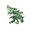
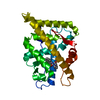
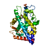
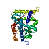
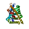
Entry Database : PDB / ID : 2ax6Title Crystal Structure Of The Androgen Receptor Ligand Binding Domain T877A Mutant In Complex With Hydroxyflutamide Androgen receptor Keywords Function / homology Function Domain/homology Component
/ / / / / / / / / / / / / / / / / / / / / / / / / / / / / / / / / / / / / / / / / / / / / / / / / / / / / / / / / / / / / / / / / / / / / / / / / / / / / / / / / / / / / / / / / / / / / / / / / / / / / / / / / / / / / / / / / / / / / / / / / / / Biological species Homo sapiens (human)Method / / Resolution : 1.5 Å Authors Bohl, C.E. / Miller, D.D. / Chen, J. / Bell, C.E. / Dalton, J.T. Journal : J.Biol.Chem. / Year : 2005Title : Structural Basis for Accommodation of Nonsteroidal Ligands in the Androgen ReceptorAuthors : Bohl, C.E. / Miller, D.D. / Chen, J. / Bell, C.E. / Dalton, J.T. History Deposition Sep 3, 2005 Deposition site / Processing site Revision 1.0 Sep 20, 2005 Provider / Type Revision 1.1 May 1, 2008 Group Revision 1.2 Jul 13, 2011 Group Revision 1.3 Oct 11, 2017 Group / Category Revision 1.4 Oct 20, 2021 Group / Derived calculations / Category / struct_ref_seq_dif / struct_siteItem _database_2.pdbx_DOI / _database_2.pdbx_database_accession ... _database_2.pdbx_DOI / _database_2.pdbx_database_accession / _struct_ref_seq_dif.details / _struct_site.pdbx_auth_asym_id / _struct_site.pdbx_auth_comp_id / _struct_site.pdbx_auth_seq_id Revision 1.5 Aug 23, 2023 Group / Refinement descriptionCategory / chem_comp_bond / pdbx_initial_refinement_model
Show all Show less
 Movie
Movie Controller

 Yorodumi
 Open data
 Basic information
 Components
 Keywords
 Function and homology information
 Homo sapiens (human)
 X-RAY DIFFRACTION /
  Authors
 Citation
 Structure visualization
 Downloads & links
 Other downloads


 PDBj
PDBj


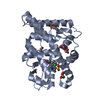
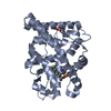
 Assembly
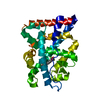

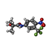
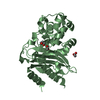
 Mass: 292.211 Da / Num. of mol.: 1 / Source method: obtained synthetically / Formula: C11H11F3N2O4
 Mass: 18.015 Da / Num. of mol.: 200 / Source method: isolated from a natural source / Formula: H2O
 Sample preparation
 Processing