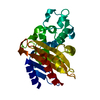
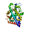
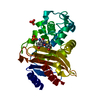
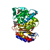
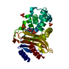
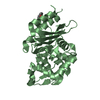
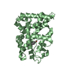
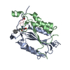
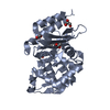
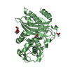
Entry Database : PDB / ID : 5oyoTitle Crystal structure of BlaC from Mycobacterium tuberculosis Beta-lactamase Keywords Function / homology Function Domain/homology Component
/ / / / / / / / / / / / / / / / Biological species Mycobacterium tuberculosis (bacteria)Method / / / Resolution : 2.1 Å Authors Tassoni, R. / Pannu, N.S. Journal : Biochemistry / Year : 2017Title : Phosphate Promotes the Recovery of Mycobacterium tuberculosis beta-Lactamase from Clavulanic Acid Inhibition.Authors : Elings, W. / Tassoni, R. / van der Schoot, S.A. / Luu, W. / Kynast, J.P. / Dai, L. / Blok, A.J. / Timmer, M. / Florea, B.I. / Pannu, N.S. / Ubbink, M. History Deposition Sep 11, 2017 Deposition site / Processing site Revision 1.0 Nov 15, 2017 Provider / Type Revision 1.1 Dec 6, 2017 Group / Category / citation_authorItem _citation.journal_volume / _citation.page_first ... _citation.journal_volume / _citation.page_first / _citation.page_last / _citation.title / _citation_author.name Revision 1.2 Jan 17, 2024 Group / Database references / Refinement descriptionCategory chem_comp_atom / chem_comp_bond ... chem_comp_atom / chem_comp_bond / database_2 / pdbx_initial_refinement_model / struct_ncs_dom_lim Item _database_2.pdbx_DOI / _database_2.pdbx_database_accession ... _database_2.pdbx_DOI / _database_2.pdbx_database_accession / _struct_ncs_dom_lim.beg_auth_comp_id / _struct_ncs_dom_lim.beg_label_asym_id / _struct_ncs_dom_lim.beg_label_comp_id / _struct_ncs_dom_lim.beg_label_seq_id / _struct_ncs_dom_lim.end_auth_comp_id / _struct_ncs_dom_lim.end_label_asym_id / _struct_ncs_dom_lim.end_label_comp_id / _struct_ncs_dom_lim.end_label_seq_id
Show all Show less
 Movie
Movie Controller


 Open data
 Basic information
 Components
 Keywords
 Function and homology information
 Mycobacterium tuberculosis (bacteria)
 X-RAY DIFFRACTION /
  Authors
 Citation
 Structure visualization
 Downloads & links
 Other downloads


 PDBj
PDBj


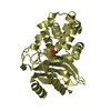
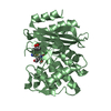
 Assembly
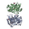

 Mass: 59.044 Da / Num. of mol.: 7 / Source method: obtained synthetically / Formula: C2H3O2
 Mass: 92.094 Da / Num. of mol.: 1 / Source method: obtained synthetically / Formula: C3H8O3
 Mass: 150.173 Da / Num. of mol.: 3 / Source method: obtained synthetically / Formula: C6H14O4
 Mass: 18.015 Da / Num. of mol.: 135 / Source method: isolated from a natural source / Formula: H2O
 Sample preparation
 / Beamline: MASSIF-1 / Wavelength: 0.96773 Å
 Processing