 Movie
Movie Controller

[English] 日本語
 Yorodumi
- PDB-5nj2: Crystal structure of BlaC from Mycobacterium tuberculosis bound t... -
+ Open data

- Basic information
| Entry | Database: PDB / ID: 5nj2 | ||||||
|---|---|---|---|---|---|---|---|
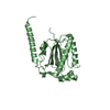
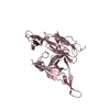
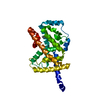
| Title | Crystal structure of BlaC from Mycobacterium tuberculosis bound to phosphate | ||||||
 Components Components | Beta-lactamase | ||||||
 Keywords Keywords | HYDROLASE | ||||||
| Function / homology |  Function and homology information Function and homology informationbeta-lactam antibiotic catabolic process / beta-lactamase activity / beta-lactamase / periplasmic space / response to antibiotic / extracellular region / plasma membrane Similarity search - Function | ||||||
| Biological species |   Mycobacterium tuberculosis (bacteria) Mycobacterium tuberculosis (bacteria) | ||||||
| Method |  X-RAY DIFFRACTION / SYNCHROTRON / MOLECULAR REPLACEMENT / Resolution: 1.19 Å X-RAY DIFFRACTION / SYNCHROTRON / MOLECULAR REPLACEMENT / Resolution: 1.19 Å | ||||||
 Authors Authors | Tassoni, R. / Pannu, N.S. / Ubbink, M. | ||||||
 Citation Citation | Journal: Biochemistry / Year: 2017 Title: Phosphate Promotes the Recovery of Mycobacterium tuberculosis beta-Lactamase from Clavulanic Acid Inhibition. Authors: Elings, W. / Tassoni, R. / van der Schoot, S.A. / Luu, W. / Kynast, J.P. / Dai, L. / Blok, A.J. / Timmer, M. / Florea, B.I. / Pannu, N.S. / Ubbink, M. | ||||||
| History |
|
- Structure visualization
| Structure viewer | Molecule: MolmilJmol/JSmol |
|---|
- Downloads & links
-Download
| PDBx/mmCIF format | 5nj2.cif.gz | 122.3 KB | Display | PDBx/mmCIF format |
|---|---|---|---|---|
| PDB format | pdb5nj2.ent.gz | 92.2 KB | Display | PDB format |
| PDBx/mmJSON format | 5nj2.json.gz | Tree view | PDBx/mmJSON format | |
| Others |  Other downloads Other downloads |
-Validation report
| Arichive directory | https://data.pdbj.org/pub/pdb/validation_reports/nj/5nj2ftp://data.pdbj.org/pub/pdb/validation_reports/nj/5nj2 | HTTPS FTP |
|---|
-Related structure data
| Related structure data |  5oyoC 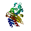 2gdnS S: Starting model for refinement C: citing same article ( |
|---|---|
| Similar structure data |


-Links
 PDBj
PDBj


- Assembly
| Deposited unit | 
| ||||||||||||||||||
|---|---|---|---|---|---|---|---|---|---|---|---|---|---|---|---|---|---|---|---|
| 1 | 
| ||||||||||||||||||
| 2 | 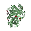
| ||||||||||||||||||
| Unit cell |
| ||||||||||||||||||
| Noncrystallographic symmetry (NCS) | NCS domain:
NCS domain segments: Component-ID: _ / Ens-ID: 1 / Beg auth comp-ID: LEU / Beg label comp-ID: LEU / End auth comp-ID: LEU / End label comp-ID: LEU / Refine code: _ / Auth seq-ID: 30 - 293 / Label seq-ID: 3 - 265
|
-Components
-Protein , 1 types, 2 molecules AB
| #1: Protein | Mass: 29475.061 Da / Num. of mol.: 2 Source method: isolated from a genetically manipulated source Source: (gene. exp.) Mycobacterium tuberculosis (bacteria) / Gene: blaC, ERS027646_02769 / Plasmid: pET28a(+) / Production host: References: UniProt: A0A0T9EA39, UniProt: P9WKD3*PLUS, beta-lactamase |
|---|
-Non-polymers , 5 types, 297 molecules 


| #2: Chemical |  Mass: 94.971 Da / Num. of mol.: 3 / Source method: obtained synthetically / Formula: PO4 Mass: 94.971 Da / Num. of mol.: 3 / Source method: obtained synthetically / Formula: PO4#3: Chemical | ChemComp-ACT /  Mass: 59.044 Da / Num. of mol.: 4 / Source method: obtained synthetically / Formula: C2H3O2 Mass: 59.044 Da / Num. of mol.: 4 / Source method: obtained synthetically / Formula: C2H3O2#4: Chemical | ChemComp-ETE / |  Mass: 208.252 Da / Num. of mol.: 1 / Source method: obtained synthetically / Formula: C9H20O5 Mass: 208.252 Da / Num. of mol.: 1 / Source method: obtained synthetically / Formula: C9H20O5#5: Chemical | ChemComp-AE4 / |  Mass: 266.331 Da / Num. of mol.: 1 / Source method: obtained synthetically / Formula: C12H26O6 Mass: 266.331 Da / Num. of mol.: 1 / Source method: obtained synthetically / Formula: C12H26O6#6: Water | ChemComp-HOH / | Mass: 18.015 Da / Num. of mol.: 288 / Source method: isolated from a natural source / Formula: H2O |
|---|
-Experimental details
-Experiment
| Experiment | Method: X-RAY DIFFRACTION / Number of used crystals: 1 |
|---|
- Sample preparation
| Crystal | Density Matthews: 2.15 Å3/Da / Density % sol: 42.88 % |
|---|---|
| Crystal grow | Temperature: 293.15 K / Method: vapor diffusion, sitting drop / Details: 0.1 M sodium acetate buffer, pH 5.0, 25% PEG1500 |
-Data collection
| Diffraction | Mean temperature: 100 K |
|---|---|
| Diffraction source | Source: SYNCHROTRON / Site: ESRF  / Beamline: MASSIF-3 / Wavelength: 0.9677 Å / Beamline: MASSIF-3 / Wavelength: 0.9677 Å |
| Detector | Type: DECTRIS EIGER X 4M / Detector: PIXEL / Date: Nov 7, 2016 |
| Radiation | Protocol: SINGLE WAVELENGTH / Monochromatic (M) / Laue (L): M / Scattering type: x-ray |
| Radiation wavelength | Wavelength: 0.9677 Å / Relative weight: 1 |
| Reflection | Resolution: 1.19→75.36 Å / Num. obs: 96841 / % possible obs: 62.7 % / Redundancy: 3.2 % / CC1/2: 0.998 / Rmerge(I) obs: 0.063 / Rpim(I) all: 0.041 / Net I/σ(I): 11.3 |
| Reflection shell | Resolution: 1.19→1.23 Å / Redundancy: 2 % / Rmerge(I) obs: 0.573 / Mean I/σ(I) obs: 1.2 / Num. unique obs: 689 / CC1/2: 0.523 / Rpim(I) all: 0.467 |
- Processing
| Software | Name: REFMAC / Version: 7.0.033 / Classification: refinement | ||||||||||||||||||||||||||||||||||||||||||||||||||||||||||||||||||||||||||||||||||||||||||||||||||||||||||||||||||||||||||||||||||||||||||||||||||||||||||||||||||||||||||||||||||||||
|---|---|---|---|---|---|---|---|---|---|---|---|---|---|---|---|---|---|---|---|---|---|---|---|---|---|---|---|---|---|---|---|---|---|---|---|---|---|---|---|---|---|---|---|---|---|---|---|---|---|---|---|---|---|---|---|---|---|---|---|---|---|---|---|---|---|---|---|---|---|---|---|---|---|---|---|---|---|---|---|---|---|---|---|---|---|---|---|---|---|---|---|---|---|---|---|---|---|---|---|---|---|---|---|---|---|---|---|---|---|---|---|---|---|---|---|---|---|---|---|---|---|---|---|---|---|---|---|---|---|---|---|---|---|---|---|---|---|---|---|---|---|---|---|---|---|---|---|---|---|---|---|---|---|---|---|---|---|---|---|---|---|---|---|---|---|---|---|---|---|---|---|---|---|---|---|---|---|---|---|---|---|---|---|
| Refinement | Method to determine structure: MOLECULAR REPLACEMENT Starting model: 2GDN Resolution: 1.19→75.36 Å / Cor.coef. Fo:Fc: 0.966 / Cor.coef. Fo:Fc free: 0.957 / SU B: 0.829 / SU ML: 0.036 / Cross valid method: THROUGHOUT / ESU R: 0.057 / ESU R Free: 0.059 / Details: HYDROGENS HAVE BEEN ADDED IN THE RIDING POSITIONS
| ||||||||||||||||||||||||||||||||||||||||||||||||||||||||||||||||||||||||||||||||||||||||||||||||||||||||||||||||||||||||||||||||||||||||||||||||||||||||||||||||||||||||||||||||||||||
| Solvent computation | Ion probe radii: 0.8 Å / Shrinkage radii: 0.8 Å / VDW probe radii: 1.2 Å | ||||||||||||||||||||||||||||||||||||||||||||||||||||||||||||||||||||||||||||||||||||||||||||||||||||||||||||||||||||||||||||||||||||||||||||||||||||||||||||||||||||||||||||||||||||||
| Displacement parameters | Biso mean: 11.803 Å2
| ||||||||||||||||||||||||||||||||||||||||||||||||||||||||||||||||||||||||||||||||||||||||||||||||||||||||||||||||||||||||||||||||||||||||||||||||||||||||||||||||||||||||||||||||||||||
| Refinement step | Cycle: 1 / Resolution: 1.19→75.36 Å
| ||||||||||||||||||||||||||||||||||||||||||||||||||||||||||||||||||||||||||||||||||||||||||||||||||||||||||||||||||||||||||||||||||||||||||||||||||||||||||||||||||||||||||||||||||||||
| Refine LS restraints |
|