 Movie
Movie Controller

[English] 日本語
 Yorodumi
- PDB-2o4r: Crystal Structure of Rat Vitamin D Receptor Ligand Binding Domain... -
+ Open data

- Basic information
| Entry | Database: PDB / ID: 2o4r | ||||||
|---|---|---|---|---|---|---|---|
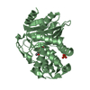
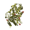
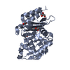
| Title | Crystal Structure of Rat Vitamin D Receptor Ligand Binding Domain Complexed with VitIII 17-20E and the NR2 Box of DRIP 205 | ||||||
 Components Components |
| ||||||
 Keywords Keywords | HORMONE/GROWTH FACTOR RECEPTOR / Nuclear receptor-ligand complex / HORMONE-GROWTH FACTOR RECEPTOR COMPLEX | ||||||
| Function / homology |  Function and homology information Function and homology informationpositive regulation of parathyroid hormone secretion / negative regulation of bone trabecula formation / Vitamin D (calciferol) metabolism / androgen biosynthetic process / regulation of RNA biosynthetic process / SUMOylation of intracellular receptors / retinal pigment epithelium development / Nuclear Receptor transcription pathway / thyroid hormone receptor signaling pathway / response to bile acid ...positive regulation of parathyroid hormone secretion / negative regulation of bone trabecula formation / Vitamin D (calciferol) metabolism / androgen biosynthetic process / regulation of RNA biosynthetic process / SUMOylation of intracellular receptors / retinal pigment epithelium development / Nuclear Receptor transcription pathway / thyroid hormone receptor signaling pathway / response to bile acid / dense fibrillar component / cellular response to vitamin D / positive regulation of apoptotic process involved in mammary gland involution / calcitriol binding / lithocholic acid binding / nuclear receptor-mediated bile acid signaling pathway / bile acid nuclear receptor activity / positive regulation of keratinocyte differentiation / ventricular trabecula myocardium morphogenesis / vitamin D binding / mediator complex / Generic Transcription Pathway / nuclear retinoic acid receptor binding / response to aldosterone / lens development in camera-type eye / cellular response to thyroid hormone stimulus / vitamin D receptor signaling pathway / positive regulation of vitamin D receptor signaling pathway / phosphate ion transmembrane transport / megakaryocyte development / negative regulation of ossification / nuclear vitamin D receptor binding / peroxisome proliferator activated receptor binding / intestinal absorption / nuclear thyroid hormone receptor binding / cellular response to steroid hormone stimulus / negative regulation of neuron differentiation / histone acetyltransferase binding / mammary gland branching involved in pregnancy / LBD domain binding / RSV-host interactions / decidualization / regulation of calcium ion transport / nuclear receptor-mediated steroid hormone signaling pathway / negative regulation of keratinocyte proliferation / keratinocyte differentiation / general transcription initiation factor binding / positive regulation of transcription initiation by RNA polymerase II / fat cell differentiation / nuclear retinoid X receptor binding / ubiquitin ligase complex / retinoic acid receptor signaling pathway / RNA polymerase II preinitiation complex assembly / erythrocyte development / intracellular receptor signaling pathway / Regulation of lipid metabolism by PPARalpha / heterochromatin / lactation / BMAL1:CLOCK,NPAS2 activates circadian expression / RORA,B,C and NR1D1 (REV-ERBA) regulate gene expression / Activation of gene expression by SREBF (SREBP) / Expression of BMAL (ARNTL), CLOCK, and NPAS2 / positive regulation of erythrocyte differentiation / T-tubule / cellular response to epidermal growth factor stimulus / animal organ morphogenesis / nuclear estrogen receptor binding / nuclear receptor binding / skeletal system development / apoptotic signaling pathway / transcription coregulator activity / Heme signaling / positive regulation of transcription elongation by RNA polymerase II / PPARA activates gene expression / promoter-specific chromatin binding / Transcriptional activation of mitochondrial biogenesis / Cytoprotection by HMOX1 / euchromatin / Nuclear Receptor transcription pathway / protein-DNA complex / Transcriptional regulation of white adipocyte differentiation / chromatin DNA binding / response to calcium ion / caveola / mRNA transcription by RNA polymerase II / cell morphogenesis / nuclear receptor activity / RNA polymerase II transcription regulator complex / transcription coactivator binding / cellular response to amyloid-beta / intracellular calcium ion homeostasis / nuclear matrix / calcium ion transport / transcription corepressor activity / response to estradiol / ubiquitin protein ligase activity / heart development / MLL4 and MLL3 complexes regulate expression of PPARG target genes in adipogenesis and hepatic steatosis / angiogenesis / Estrogen-dependent gene expression Similarity search - Function | ||||||
| Biological species |  | ||||||
| Method |  X-RAY DIFFRACTION / FOURIER SYNTHESIS / Resolution: 1.98 Å X-RAY DIFFRACTION / FOURIER SYNTHESIS / Resolution: 1.98 Å | ||||||
 Authors Authors | Vanhooke, J.L. / Benning, M.M. / DeLuca, H.F. | ||||||
 Citation Citation | Journal: Arch.Biochem.Biophys. / Year: 2007 Title: New analogs of 2-methylene-19-nor-(20S)-1,25-dihydroxyvitamin D(3) with conformationally restricted side chains: Evaluation of biological activity and structural determination of VDR-bound conformations. Authors: Vanhooke, J.L. / Tadi, B.P. / Benning, M.M. / Plum, L.A. / Deluca, H.F. #1: Journal: Biochemistry / Year: 2004Title: Molecular Structure of the Rat Vitamin D Receptor Ligand Binding Domain Complexed with 2-Carbon-Substituted Vitamin D3 Hormone Analogues and a LXXLL-Containing Coactivator Peptide Authors: Vanhooke, J.L. / Benning, M.M. / Bauer, C.B. / Pike, J.W. / DeLuca, H.F. | ||||||
| History |
|
- Structure visualization
| Structure viewer | Molecule: MolmilJmol/JSmol |
|---|
- Downloads & links
-Download
| PDBx/mmCIF format | 2o4r.cif.gz | 71.5 KB | Display | PDBx/mmCIF format |
|---|---|---|---|---|
| PDB format | pdb2o4r.ent.gz | 50.7 KB | Display | PDB format |
| PDBx/mmJSON format | 2o4r.json.gz | Tree view | PDBx/mmJSON format | |
| Others |  Other downloads Other downloads |
-Validation report
| Arichive directory | https://data.pdbj.org/pub/pdb/validation_reports/o4/2o4rftp://data.pdbj.org/pub/pdb/validation_reports/o4/2o4r | HTTPS FTP |
|---|
-Related structure data
| Related structure data |  2o4jC  1rjkS S: Starting model for refinement C: citing same article ( |
|---|---|
| Similar structure data |


-Links
 PDBj
PDBj


- Assembly
| Deposited unit | 
| ||||||||
|---|---|---|---|---|---|---|---|---|---|
| 1 |
| ||||||||
| 2 | 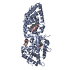
| ||||||||
| Unit cell |
| ||||||||
| Details | The biological unit is composed of one molecule of VDR (chain A), one molecule of the synthetic peptide (chain C), and one ligand molecule (residue name VD5). The asymmetric unit contains one biological unit. |
-Components
| #1: Protein | Mass: 32983.730 Da / Num. of mol.: 1 / Fragment: ligand binding domain Mutation: residues Ser165 through Pro211 are deleted from the protein Source method: isolated from a genetically manipulated source Source: (gene. exp.)  |
|---|---|
| #2: Protein/peptide | Mass: 1570.898 Da / Num. of mol.: 1 / Fragment: DRIP 205 NR2 Box Peptide / Source method: obtained synthetically / Details: synthesized at UW-Madison Biotechnology Center / References: UniProt: Q15648 |
| #3: Chemical | ChemComp-VD5 / (  Mass: 414.621 Da / Num. of mol.: 1 / Source method: obtained synthetically / Formula: C27H42O3 Mass: 414.621 Da / Num. of mol.: 1 / Source method: obtained synthetically / Formula: C27H42O3 |
| #4: Water | ChemComp-HOH /  Mass: 18.015 Da / Num. of mol.: 166 / Source method: isolated from a natural source / Formula: H2O Mass: 18.015 Da / Num. of mol.: 166 / Source method: isolated from a natural source / Formula: H2O |
-Experimental details
-Experiment
| Experiment | Method: X-RAY DIFFRACTION / Number of used crystals: 1 |
|---|
- Sample preparation
| Crystal | Density Matthews: 2 Å3/Da / Density % sol: 38.5 % |
|---|---|
| Crystal grow | Temperature: 295 K / Method: macroseeding in batch / pH: 7 Details: PEG 4000, MOPS, Ammonium Citrate, Isopropanol, pH 7.0, macroseeding in batch, temperature 295K |
-Data collection
| Diffraction | Mean temperature: 100 K |
|---|---|
| Diffraction source | Source: ROTATING ANODE / Wavelength: 1.5418 |
| Detector | Type: BRUKER PROTEUM R / Detector: CCD / Date: Nov 30, 2005 / Details: Montel Optics |
| Radiation | Protocol: SINGLE WAVELENGTH / Scattering type: x-ray |
| Radiation wavelength | Wavelength: 1.5418 Å / Relative weight: 1 |
| Reflection | Resolution: 1.98→42.5 Å / Num. all: 19228 / Num. obs: 19195 / % possible obs: 99.8 % / Observed criterion σ(F): 0 / Observed criterion σ(I): 3 / Redundancy: 7.03 % / Rsym value: 0.0447 / Net I/σ(I): 22.77 |
| Reflection shell | Resolution: 1.98→2.08 Å / Redundancy: 3.61 % / Mean I/σ(I) obs: 4.49 / Num. unique all: 2611 / Rsym value: 0.186 / % possible all: 99.6 |
- Processing
| Software |
| ||||||||||||||||||||||||||||||||||||||||||||||||||||||||||||||||||||||||||||||||||||||||||
|---|---|---|---|---|---|---|---|---|---|---|---|---|---|---|---|---|---|---|---|---|---|---|---|---|---|---|---|---|---|---|---|---|---|---|---|---|---|---|---|---|---|---|---|---|---|---|---|---|---|---|---|---|---|---|---|---|---|---|---|---|---|---|---|---|---|---|---|---|---|---|---|---|---|---|---|---|---|---|---|---|---|---|---|---|---|---|---|---|---|---|---|
| Refinement | Method to determine structure: FOURIER SYNTHESIS Starting model: pdb entry 1RJK Resolution: 1.98→30 Å / Cor.coef. Fo:Fc: 0.957 / Cor.coef. Fo:Fc free: 0.929 / SU B: 4.149 / SU ML: 0.12 / Cross valid method: THROUGHOUT / σ(F): 0 / ESU R: 0.189 / ESU R Free: 0.173 / Stereochemistry target values: MAXIMUM LIKELIHOOD / Details: HYDROGENS HAVE BEEN ADDED IN THE RIDING POSITIONS
| ||||||||||||||||||||||||||||||||||||||||||||||||||||||||||||||||||||||||||||||||||||||||||
| Solvent computation | Ion probe radii: 0.8 Å / Shrinkage radii: 0.8 Å / VDW probe radii: 1.4 Å / Solvent model: MASK | ||||||||||||||||||||||||||||||||||||||||||||||||||||||||||||||||||||||||||||||||||||||||||
| Displacement parameters | Biso mean: 26.771 Å2
| ||||||||||||||||||||||||||||||||||||||||||||||||||||||||||||||||||||||||||||||||||||||||||
| Refinement step | Cycle: LAST / Resolution: 1.98→30 Å
| ||||||||||||||||||||||||||||||||||||||||||||||||||||||||||||||||||||||||||||||||||||||||||
| Refine LS restraints |
| ||||||||||||||||||||||||||||||||||||||||||||||||||||||||||||||||||||||||||||||||||||||||||
| LS refinement shell | Resolution: 1.98→2.031 Å / Total num. of bins used: 20
|
