 Movie
Movie Controller

[English] 日本語
 Yorodumi
- PDB-1rk3: crystal structure of the rat vitamin D receptor ligand binding do... -
+ Open data

- Basic information
| Entry | Database: PDB / ID: 1rk3 | ||||||
|---|---|---|---|---|---|---|---|
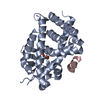
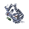
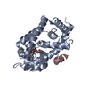
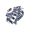
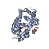
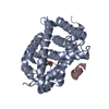
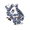
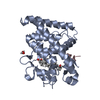
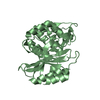
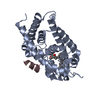
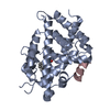
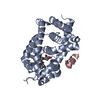
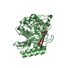
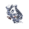
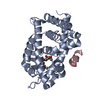
| Title | crystal structure of the rat vitamin D receptor ligand binding domain complexed with 1,25-dihydroxyvitamin D3 and a synthetic peptide containing the NR2 box of DRIP 205 | ||||||
 Components Components |
| ||||||
 Keywords Keywords | hormone/growth factor receptor / nuclear receptor-ligand complex / nuclear receptor-coactivator interactions / hormone-growth factor receptor COMPLEX | ||||||
| Function / homology |  Function and homology information Function and homology informationnegative regulation of bone trabecula formation / Vitamin D (calciferol) metabolism / negative regulation of phosphate transmembrane transport / enucleate erythrocyte development / positive regulation of type II interferon-mediated signaling pathway / androgen biosynthetic process / positive regulation of G0 to G1 transition / regulation of RNA biosynthetic process / SUMOylation of intracellular receptors / retinal pigment epithelium development ...negative regulation of bone trabecula formation / Vitamin D (calciferol) metabolism / negative regulation of phosphate transmembrane transport / enucleate erythrocyte development / positive regulation of type II interferon-mediated signaling pathway / androgen biosynthetic process / positive regulation of G0 to G1 transition / regulation of RNA biosynthetic process / SUMOylation of intracellular receptors / retinal pigment epithelium development / G0 to G1 transition / Nuclear Receptor transcription pathway / thyroid hormone receptor signaling pathway / response to bile acid / mammary gland branching involved in thelarche / dense fibrillar component / core mediator complex / positive regulation of parathyroid hormone secretion / cell-cell junction maintenance / regulation of vitamin D receptor signaling pathway / positive regulation of apoptotic process involved in mammary gland involution / calcitriol binding / cellular response to vitamin D / lithocholic acid binding / nuclear receptor-mediated bile acid signaling pathway / bile acid nuclear receptor activity / ventricular trabecula myocardium morphogenesis / positive regulation of hepatocyte proliferation / positive regulation of keratinocyte differentiation / vitamin D binding / mediator complex / thyroid hormone generation / Generic Transcription Pathway / nuclear retinoic acid receptor binding / embryonic heart tube development / response to aldosterone / cellular response to thyroid hormone stimulus / vitamin D receptor signaling pathway / positive regulation of vitamin D receptor signaling pathway / phosphate ion transmembrane transport / embryonic hindlimb morphogenesis / lens development in camera-type eye / nuclear vitamin D receptor binding / negative regulation of ossification / peroxisome proliferator activated receptor binding / intestinal absorption / embryonic hemopoiesis / nuclear thyroid hormone receptor binding / megakaryocyte development / cellular response to hepatocyte growth factor stimulus / cellular response to steroid hormone stimulus / positive regulation of intracellular estrogen receptor signaling pathway / negative regulation of neuron differentiation / epithelial cell proliferation involved in mammary gland duct elongation / histone acetyltransferase binding / LBD domain binding / mammary gland branching involved in pregnancy / RSV-host interactions / nuclear steroid receptor activity / decidualization / regulation of calcium ion transport / nuclear receptor-mediated steroid hormone signaling pathway / animal organ regeneration / negative regulation of keratinocyte proliferation / monocyte differentiation / general transcription initiation factor binding / embryonic placenta development / hematopoietic stem cell differentiation / positive regulation of transcription initiation by RNA polymerase II / nuclear retinoid X receptor binding / fat cell differentiation / ubiquitin ligase complex / RNA polymerase II preinitiation complex assembly / retinoic acid receptor signaling pathway / keratinocyte differentiation / erythrocyte development / intracellular receptor signaling pathway / Regulation of lipid metabolism by PPARalpha / lactation / heterochromatin / peroxisome proliferator activated receptor signaling pathway / BMAL1:CLOCK,NPAS2 activates circadian expression / RORA,B,C and NR1D1 (REV-ERBA) regulate gene expression / Activation of gene expression by SREBF (SREBP) / Expression of BMAL (ARNTL), CLOCK, and NPAS2 / T-tubule / positive regulation of erythrocyte differentiation / cellular response to epidermal growth factor stimulus / nuclear estrogen receptor binding / nuclear receptor binding / animal organ morphogenesis / skeletal system development / transcription coregulator activity / Heme signaling / positive regulation of transcription elongation by RNA polymerase II / liver development / promoter-specific chromatin binding / apoptotic signaling pathway / PPARA activates gene expression / Transcriptional activation of mitochondrial biogenesis Similarity search - Function | ||||||
| Biological species |  | ||||||
| Method |  X-RAY DIFFRACTION / FOURIER SYNTHESIS / Resolution: 2.2 Å X-RAY DIFFRACTION / FOURIER SYNTHESIS / Resolution: 2.2 Å | ||||||
 Authors Authors | Vanhooke, J.L. / M Benning, M. / Bauer, C.B. / Pike, J.W. / DeLuca, H.F. | ||||||
 Citation Citation | Journal: Biochemistry / Year: 2004 Title: Molecular Structure of the Rat Vitamin D Receptor Ligand Binding Domain Complexed with 2-Carbon-Substituted Vitamin D(3) Hormone Analogues and a LXXLL-Containing Coactivator Peptide Authors: Vanhooke, J.L. / Benning, M.M. / Bauer, C.B. / Pike, J.W. / DeLuca, H.F. | ||||||
| History |
|
- Structure visualization
| Structure viewer | Molecule: MolmilJmol/JSmol |
|---|
- Downloads & links
-Download
| PDBx/mmCIF format | 1rk3.cif.gz | 68.6 KB | Display | PDBx/mmCIF format |
|---|---|---|---|---|
| PDB format | pdb1rk3.ent.gz | 48.8 KB | Display | PDB format |
| PDBx/mmJSON format | 1rk3.json.gz | Tree view | PDBx/mmJSON format | |
| Others |  Other downloads Other downloads |
-Validation report
| Arichive directory | https://data.pdbj.org/pub/pdb/validation_reports/rk/1rk3ftp://data.pdbj.org/pub/pdb/validation_reports/rk/1rk3 | HTTPS FTP |
|---|
-Related structure data
| Related structure data |  1rjkSC  1rkgC  1rkhC S: Starting model for refinement C: citing same article ( |
|---|---|
| Similar structure data |


-Links
 PDBj
PDBj


- Assembly
| Deposited unit | 
| ||||||||
|---|---|---|---|---|---|---|---|---|---|
| 1 |
| ||||||||
| 2 | 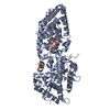
| ||||||||
| Unit cell |
| ||||||||
| Details | The asymmetric unit contains one biological unit. The biological unit is composed of one molecule of VDR, one ligand molecule, and one molecule of the synthetic peptide |
-Components
| #1: Protein | Mass: 32983.730 Da / Num. of mol.: 1 / Fragment: ligand binding domain / Mutation: Chain A, DEL(165-211) Source method: isolated from a genetically manipulated source Source: (gene. exp.)  |
|---|---|
| #2: Protein/peptide | Mass: 1570.898 Da / Num. of mol.: 1 / Fragment: DRIP 205 NR2 box peptide / Source method: obtained synthetically / References: UniProt: Q15648 |
| #3: Chemical | ChemComp-VDX /   Mass: 416.636 Da / Num. of mol.: 1 / Source method: obtained synthetically / Formula: C27H44O3 / Comment: hormone*YM Mass: 416.636 Da / Num. of mol.: 1 / Source method: obtained synthetically / Formula: C27H44O3 / Comment: hormone*YM |
| #4: Water | ChemComp-HOH /  Mass: 18.015 Da / Num. of mol.: 92 / Source method: isolated from a natural source / Formula: H2O Mass: 18.015 Da / Num. of mol.: 92 / Source method: isolated from a natural source / Formula: H2O |
-Experimental details
-Experiment
| Experiment | Method: X-RAY DIFFRACTION / Number of used crystals: 1 |
|---|
- Sample preparation
| Crystal | Density Matthews: 2.07 Å3/Da / Density % sol: 40.48 % |
|---|---|
| Crystal grow | Temperature: 295 K / Method: vapor diffusion, hanging drop / pH: 7 Details: PEG 4000, MOPS, ammonium citrate, isopropanol, pH 7.0, VAPOR DIFFUSION, HANGING DROP, temperature 295K |
-Data collection
| Diffraction | Mean temperature: 100 K |
|---|---|
| Diffraction source | Source: ROTATING ANODE / Type: ENRAF-NONIUS FR591 / Wavelength: 1.5418 Å |
| Detector | Type: BRUKER PROTEUM R / Detector: CCD / Date: Oct 24, 2002 |
| Radiation | Monochromator: Montel optics / Protocol: SINGLE WAVELENGTH / Monochromatic (M) / Laue (L): M / Scattering type: x-ray |
| Radiation wavelength | Wavelength: 1.5418 Å / Relative weight: 1 |
| Reflection | Resolution: 2.2→42.44 Å / Num. all: 14565 / Num. obs: 14475 / % possible obs: 99 % / Observed criterion σ(F): 0 / Observed criterion σ(I): 3 / Redundancy: 5.7 % / Rsym value: 0.052 / Net I/σ(I): 10.9 |
| Reflection shell | Resolution: 2.2→2.3 Å / Mean I/σ(I) obs: 2.7 / Rsym value: 0.198 / % possible all: 95 |
- Processing
| Software |
| ||||||||||||||||||||||||||||||||||||||||||||||||||||||||||||||||||||||
|---|---|---|---|---|---|---|---|---|---|---|---|---|---|---|---|---|---|---|---|---|---|---|---|---|---|---|---|---|---|---|---|---|---|---|---|---|---|---|---|---|---|---|---|---|---|---|---|---|---|---|---|---|---|---|---|---|---|---|---|---|---|---|---|---|---|---|---|---|---|---|---|
| Refinement | Method to determine structure: FOURIER SYNTHESIS Starting model: PDB entry 1RJK Resolution: 2.2→30 Å / Cor.coef. Fo:Fc: 0.95 / Cor.coef. Fo:Fc free: 0.919 / SU B: 5.508 / SU ML: 0.142 / Cross valid method: THROUGHOUT / σ(F): 0 / ESU R: 0.27 / ESU R Free: 0.213 / Stereochemistry target values: MAXIMUM LIKELIHOOD
| ||||||||||||||||||||||||||||||||||||||||||||||||||||||||||||||||||||||
| Solvent computation | Ion probe radii: 0.8 Å / Shrinkage radii: 0.8 Å / VDW probe radii: 1.4 Å / Solvent model: BABINET MODEL WITH MASK | ||||||||||||||||||||||||||||||||||||||||||||||||||||||||||||||||||||||
| Displacement parameters | Biso mean: 34.7 Å2
| ||||||||||||||||||||||||||||||||||||||||||||||||||||||||||||||||||||||
| Refinement step | Cycle: LAST / Resolution: 2.2→30 Å
| ||||||||||||||||||||||||||||||||||||||||||||||||||||||||||||||||||||||
| Refine LS restraints |
| ||||||||||||||||||||||||||||||||||||||||||||||||||||||||||||||||||||||
| LS refinement shell | Resolution: 2.201→2.258 Å / Total num. of bins used: 20
|
