 Movie
Movie Controller

[English] 日本語
 Yorodumi
- PDB-1or7: Crystal Structure of Escherichia coli sigmaE with the Cytoplasmic... -
+ Open data

- Basic information
| Entry | Database: PDB / ID: 1or7 | ||||||
|---|---|---|---|---|---|---|---|
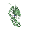
| Title | Crystal Structure of Escherichia coli sigmaE with the Cytoplasmic Domain of its Anti-sigma RseA | ||||||
 Components Components |
| ||||||
 Keywords Keywords | TRANSCRIPTION / regulation / DNA-BINDING / TRANSMEMBRANE | ||||||
| Function / homology |  Function and homology information Function and homology informationsigma factor antagonist activity / sigma factor antagonist complex / response to osmotic stress / response to temperature stimulus / submerged biofilm formation / cellular response to cell envelope stress / regulation of DNA-templated transcription initiation / response to stress / sigma factor activity / cytosolic DNA-directed RNA polymerase complex ...sigma factor antagonist activity / sigma factor antagonist complex / response to osmotic stress / response to temperature stimulus / submerged biofilm formation / cellular response to cell envelope stress / regulation of DNA-templated transcription initiation / response to stress / sigma factor activity / cytosolic DNA-directed RNA polymerase complex / DNA-templated transcription initiation / molecular adaptor activity / negative regulation of DNA-templated transcription / regulation of DNA-templated transcription / DNA-templated transcription / DNA binding / plasma membrane / cytoplasm Similarity search - Function | ||||||
| Biological species |  | ||||||
| Method |  X-RAY DIFFRACTION / SYNCHROTRON / MAD / Resolution: 2 Å X-RAY DIFFRACTION / SYNCHROTRON / MAD / Resolution: 2 Å | ||||||
 Authors Authors | Campbell, E.A. / Tupy, J.L. / Gruber, T.M. / Wang, S. / Sharp, M.M. / Gross, C.A. / Darst, S.A. | ||||||
 Citation Citation | Journal: Mol.Cell / Year: 2003 Title: Crystal structure of Escherichia coli sigmaE with the cytoplasmic domain of its anti-sigma RseA. Authors: Campbell, E.A. / Tupy, J.L. / Gruber, T.M. / Wang, S. / Sharp, M.M. / Gross, C.A. / Darst, S.A. | ||||||
| History |
|
- Structure visualization
| Structure viewer | Molecule: MolmilJmol/JSmol |
|---|
- Downloads & links
-Download
| PDBx/mmCIF format | 1or7.cif.gz | 111.1 KB | Display | PDBx/mmCIF format |
|---|---|---|---|---|
| PDB format | pdb1or7.ent.gz | 87.1 KB | Display | PDB format |
| PDBx/mmJSON format | 1or7.json.gz | Tree view | PDBx/mmJSON format | |
| Others |  Other downloads Other downloads |
-Validation report
| Arichive directory | https://data.pdbj.org/pub/pdb/validation_reports/or/1or7ftp://data.pdbj.org/pub/pdb/validation_reports/or/1or7 | HTTPS FTP |
|---|
-Related structure data
| Similar structure data |
|---|


-Links
 PDBj
PDBj
- Assembly
| Deposited unit | 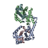
| ||||||||
|---|---|---|---|---|---|---|---|---|---|
| 1 | 
| ||||||||
| 2 | 
| ||||||||
| 3 |
| ||||||||
| Unit cell |
| ||||||||
| Details | The biological assembly is a heterodimer of sigma E (chains A,B) plus RseA (chain C), or chains (D,E) plus chain F |
-Components
| #1: Protein | Mass: 22003.986 Da / Num. of mol.: 2 Source method: isolated from a genetically manipulated source Source: (gene. exp.) Gene: RPOE OR SIGE OR B2573 OR C3097 OR Z3855 OR ECS3439 OR SF2635 Plasmid: pLC31 / Species (production host): Escherichia coli / Production host: #2: Protein | Mass: 10408.719 Da / Num. of mol.: 2 / Fragment: RseA-N, residues 1-90 Source method: isolated from a genetically manipulated source Source: (gene. exp.) #3: Water | ChemComp-HOH / |  Mass: 18.015 Da / Num. of mol.: 175 / Source method: isolated from a natural source / Formula: H2O Mass: 18.015 Da / Num. of mol.: 175 / Source method: isolated from a natural source / Formula: H2O |
|---|
-Experimental details
-Experiment
| Experiment | Method: X-RAY DIFFRACTION / Number of used crystals: 1 |
|---|
- Sample preparation
| Crystal | Density Matthews: 2.82 Å3/Da / Density % sol: 56.04 % | ||||||||||||||||||||||||
|---|---|---|---|---|---|---|---|---|---|---|---|---|---|---|---|---|---|---|---|---|---|---|---|---|---|
| Crystal grow | Temperature: 277 K / Method: vapor diffusion, hanging drop / pH: 6 Details: sodium formate, MES buffer, pH 6, VAPOR DIFFUSION, HANGING DROP, temperature 277K | ||||||||||||||||||||||||
| Crystal grow | *PLUS Temperature: 4 ℃ / pH: 6 / Method: vapor diffusion | ||||||||||||||||||||||||
| Components of the solutions | *PLUS
|
-Data collection
| Diffraction | Mean temperature: 100 K |
|---|---|
| Diffraction source | Source: SYNCHROTRON / Site: NSLS  / Beamline: X9A / Wavelength: 0.9792 Å / Beamline: X9A / Wavelength: 0.9792 Å |
| Detector | Type: MARRESEARCH / Detector: CCD / Date: Apr 26, 2002 |
| Radiation | Monochromator: Si 111 / Protocol: MAD / Monochromatic (M) / Laue (L): M / Scattering type: x-ray |
| Radiation wavelength | Wavelength: 0.9792 Å / Relative weight: 1 |
| Reflection | Resolution: 2→35 Å / Num. all: 84266 / Num. obs: 82833 / % possible obs: 98.3 % / Observed criterion σ(F): -3 / Observed criterion σ(I): -3 / Redundancy: 2.2 % / Rsym value: 0.083 / Net I/σ(I): 16.4 |
| Reflection shell | Resolution: 2→2.07 Å / Redundancy: 3 % / Mean I/σ(I) obs: 3.6 / Num. unique all: 4296 / Rsym value: 0.359 / % possible all: 92.3 |
| Reflection | *PLUS Highest resolution: 2 Å / Lowest resolution: 35 Å / Num. measured all: 181227 / Rmerge(I) obs: 0.069 |
| Reflection shell | *PLUS % possible obs: 92.8 % / Rmerge(I) obs: 0.381 / Mean I/σ(I) obs: 2.6 |
- Processing
| Software |
| |||||||||||||||||||||||||||||||||||||||||||||||||||||||||||||||||||||||||||||||||||||||||||||||||||||||||||||||||||||||||||||||||||||||||||||||||||||||||||||||||||||||||||||||
|---|---|---|---|---|---|---|---|---|---|---|---|---|---|---|---|---|---|---|---|---|---|---|---|---|---|---|---|---|---|---|---|---|---|---|---|---|---|---|---|---|---|---|---|---|---|---|---|---|---|---|---|---|---|---|---|---|---|---|---|---|---|---|---|---|---|---|---|---|---|---|---|---|---|---|---|---|---|---|---|---|---|---|---|---|---|---|---|---|---|---|---|---|---|---|---|---|---|---|---|---|---|---|---|---|---|---|---|---|---|---|---|---|---|---|---|---|---|---|---|---|---|---|---|---|---|---|---|---|---|---|---|---|---|---|---|---|---|---|---|---|---|---|---|---|---|---|---|---|---|---|---|---|---|---|---|---|---|---|---|---|---|---|---|---|---|---|---|---|---|---|---|---|---|---|---|---|
| Refinement | Method to determine structure: MAD / Resolution: 2→30 Å / Cor.coef. Fo:Fc: 0.948 / Cor.coef. Fo:Fc free: 0.929 / SU B: 5.101 / SU ML: 0.143 / TLS residual ADP flag: LIKELY RESIDUAL / Cross valid method: THROUGHOUT / σ(F): -3 / ESU R: 0.162 / ESU R Free: 0.148 Stereochemistry target values: MAXIMUM LIKELIHOOD WITH PHASES Details: HYDROGENS HAVE BEEN ADDED IN THE RIDING POSITIONS
| |||||||||||||||||||||||||||||||||||||||||||||||||||||||||||||||||||||||||||||||||||||||||||||||||||||||||||||||||||||||||||||||||||||||||||||||||||||||||||||||||||||||||||||||
| Solvent computation | Ion probe radii: 0.8 Å / Shrinkage radii: 0.8 Å / VDW probe radii: 1.4 Å / Solvent model: BABINET MODEL WITH MASK | |||||||||||||||||||||||||||||||||||||||||||||||||||||||||||||||||||||||||||||||||||||||||||||||||||||||||||||||||||||||||||||||||||||||||||||||||||||||||||||||||||||||||||||||
| Displacement parameters | Biso mean: 19.358 Å2
| |||||||||||||||||||||||||||||||||||||||||||||||||||||||||||||||||||||||||||||||||||||||||||||||||||||||||||||||||||||||||||||||||||||||||||||||||||||||||||||||||||||||||||||||
| Refinement step | Cycle: LAST / Resolution: 2→30 Å
| |||||||||||||||||||||||||||||||||||||||||||||||||||||||||||||||||||||||||||||||||||||||||||||||||||||||||||||||||||||||||||||||||||||||||||||||||||||||||||||||||||||||||||||||
| Refine LS restraints |
| |||||||||||||||||||||||||||||||||||||||||||||||||||||||||||||||||||||||||||||||||||||||||||||||||||||||||||||||||||||||||||||||||||||||||||||||||||||||||||||||||||||||||||||||
| LS refinement shell | Resolution: 2→2.052 Å / Total num. of bins used: 20 /
| |||||||||||||||||||||||||||||||||||||||||||||||||||||||||||||||||||||||||||||||||||||||||||||||||||||||||||||||||||||||||||||||||||||||||||||||||||||||||||||||||||||||||||||||
| Refinement TLS params. | Method: refined / Refine-ID: X-RAY DIFFRACTION
| |||||||||||||||||||||||||||||||||||||||||||||||||||||||||||||||||||||||||||||||||||||||||||||||||||||||||||||||||||||||||||||||||||||||||||||||||||||||||||||||||||||||||||||||
| Refinement TLS group |
| |||||||||||||||||||||||||||||||||||||||||||||||||||||||||||||||||||||||||||||||||||||||||||||||||||||||||||||||||||||||||||||||||||||||||||||||||||||||||||||||||||||||||||||||
| Refinement | *PLUS Highest resolution: 2 Å / Lowest resolution: 30 Å / % reflection Rfree: 10 % / Rfactor Rfree: 0.232 / Rfactor Rwork: 0.197 | |||||||||||||||||||||||||||||||||||||||||||||||||||||||||||||||||||||||||||||||||||||||||||||||||||||||||||||||||||||||||||||||||||||||||||||||||||||||||||||||||||||||||||||||
| Solvent computation | *PLUS | |||||||||||||||||||||||||||||||||||||||||||||||||||||||||||||||||||||||||||||||||||||||||||||||||||||||||||||||||||||||||||||||||||||||||||||||||||||||||||||||||||||||||||||||
| Displacement parameters | *PLUS | |||||||||||||||||||||||||||||||||||||||||||||||||||||||||||||||||||||||||||||||||||||||||||||||||||||||||||||||||||||||||||||||||||||||||||||||||||||||||||||||||||||||||||||||
| Refine LS restraints | *PLUS
|
