 Movie
Movie Controller

[English] 日本語
 Yorodumi
- PDB-3qy2: Crystal structure of the P93A monomer mutant of S. cerevisiae Cks1 -
+ Open data

- Basic information
| Entry | Database: PDB / ID: 3qy2 | ||||||
|---|---|---|---|---|---|---|---|
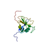
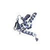
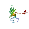
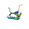
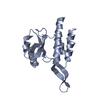
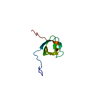
| Title | Crystal structure of the P93A monomer mutant of S. cerevisiae Cks1 | ||||||
 Components Components | Cyclin-dependent kinases regulatory subunit | ||||||
 Keywords Keywords | TRANSFERASE REGULATOR / protein kinase activator / ubiquitin binding / transcription / Cdc28 kinase / cell cycle / cyclin-dependent kinase | ||||||
| Function / homology |  Function and homology information Function and homology informationregulation of cell cycle G1/S phase transition / cyclin-dependent protein serine/threonine kinase activator activity / SCF ubiquitin ligase complex / protein kinase activator activity / cyclin-dependent protein kinase holoenzyme complex / regulation of mitotic cell cycle / ubiquitin binding / histone binding / cell division / regulation of transcription by RNA polymerase II ...regulation of cell cycle G1/S phase transition / cyclin-dependent protein serine/threonine kinase activator activity / SCF ubiquitin ligase complex / protein kinase activator activity / cyclin-dependent protein kinase holoenzyme complex / regulation of mitotic cell cycle / ubiquitin binding / histone binding / cell division / regulation of transcription by RNA polymerase II / protein kinase binding / positive regulation of transcription by RNA polymerase II / nucleus / cytoplasm Similarity search - Function | ||||||
| Biological species |  | ||||||
| Method |  X-RAY DIFFRACTION / SYNCHROTRON / MOLECULAR REPLACEMENT / Resolution: 2.59 Å X-RAY DIFFRACTION / SYNCHROTRON / MOLECULAR REPLACEMENT / Resolution: 2.59 Å | ||||||
 Authors Authors | Balog, E.R.M. / Saetern, O.C. / Finch, W. / Hoeft, C.O. / Thai, V. / Harvey, S.L. / Kellogg, D.K. / Rubin, S.M. | ||||||
 Citation Citation | Journal: J.Mol.Biol. / Year: 2011 Title: The structure of a monomeric mutant cks protein reveals multiple functions for a conserved hinge-region proline. Authors: Balog, E.R. / Saetern, O.C. / Finch, W. / Hoeft, C.O. / Thai, V. / Harvey, S.L. / Kellogg, D.R. / Rubin, S.M. | ||||||
| History |
|
- Structure visualization
| Structure viewer | Molecule: MolmilJmol/JSmol |
|---|
- Downloads & links
-Download
| PDBx/mmCIF format | 3qy2.cif.gz | 56.7 KB | Display | PDBx/mmCIF format |
|---|---|---|---|---|
| PDB format | pdb3qy2.ent.gz | 41.9 KB | Display | PDB format |
| PDBx/mmJSON format | 3qy2.json.gz | Tree view | PDBx/mmJSON format | |
| Others |  Other downloads Other downloads |
-Validation report
| Arichive directory | https://data.pdbj.org/pub/pdb/validation_reports/qy/3qy2ftp://data.pdbj.org/pub/pdb/validation_reports/qy/3qy2 | HTTPS FTP |
|---|
-Related structure data
| Similar structure data |
|---|


-Links
 PDBj
PDBj


- Assembly
| Deposited unit | 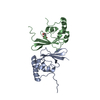
| ||||||||
|---|---|---|---|---|---|---|---|---|---|
| 1 | 
| ||||||||
| 2 | 
| ||||||||
| 3 | 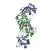
| ||||||||
| 4 | 
| ||||||||
| Unit cell |
| ||||||||
| Components on special symmetry positions |
|
-Components
| #1: Protein | Mass: 13847.542 Da / Num. of mol.: 2 / Fragment: UNP residues 1-117 / Mutation: P93A Source method: isolated from a genetically manipulated source Source: (gene. exp.) Gene: CKS1, YBR135W, YBR1011 / Production host:  #2: Chemical | ChemComp-FLC / |   Mass: 189.100 Da / Num. of mol.: 1 / Source method: obtained synthetically / Formula: C6H5O7 Mass: 189.100 Da / Num. of mol.: 1 / Source method: obtained synthetically / Formula: C6H5O7#3: Water | ChemComp-HOH / |  Mass: 18.015 Da / Num. of mol.: 42 / Source method: isolated from a natural source / Formula: H2O Mass: 18.015 Da / Num. of mol.: 42 / Source method: isolated from a natural source / Formula: H2O |
|---|
-Experimental details
-Experiment
| Experiment | Method: X-RAY DIFFRACTION / Number of used crystals: 1 |
|---|
- Sample preparation
| Crystal | Density Matthews: 3.37 Å3/Da / Density % sol: 63.5 % |
|---|---|
| Crystal grow | Temperature: 298 K / Method: vapor diffusion, hanging drop / pH: 6.5 Details: 0.1 M sodium cacodylate, 0.2 M sodium citrate, 5% isopropanol, pH 6.5, VAPOR DIFFUSION, HANGING DROP, temperature 298K |
-Data collection
| Diffraction | Mean temperature: 100 K |
|---|---|
| Diffraction source | Source: SYNCHROTRON / Site: ALS  / Beamline: 5.0.1 / Wavelength: 0.9774 Å / Beamline: 5.0.1 / Wavelength: 0.9774 Å |
| Detector | Type: ADSC QUANTUM 315r / Detector: CCD / Date: Oct 23, 2009 |
| Radiation | Monochromator: Si(220) Asymmetric cut single crystal / Protocol: SINGLE WAVELENGTH / Monochromatic (M) / Laue (L): M / Scattering type: x-ray |
| Radiation wavelength | Wavelength: 0.9774 Å / Relative weight: 1 |
| Reflection | Resolution: 2.6→50 Å / Num. obs: 12295 / % possible obs: 100 % / Observed criterion σ(F): 0 / Observed criterion σ(I): -3 / Biso Wilson estimate: 9.4 Å2 / Rmerge(I) obs: 0.081 / Rsym value: 0.081 / Net I/σ(I): 31.167 |
| Reflection shell | Resolution: 2.6→2.64 Å / Redundancy: 9.3 % / Rmerge(I) obs: 0.41 / Mean I/σ(I) obs: 5.417 / Rsym value: 0.41 / % possible all: 100 |
- Processing
| Software |
| |||||||||||||||||||||||||||||||||||||||||||||||||||||||||||||||||
|---|---|---|---|---|---|---|---|---|---|---|---|---|---|---|---|---|---|---|---|---|---|---|---|---|---|---|---|---|---|---|---|---|---|---|---|---|---|---|---|---|---|---|---|---|---|---|---|---|---|---|---|---|---|---|---|---|---|---|---|---|---|---|---|---|---|---|
| Refinement | Method to determine structure: MOLECULAR REPLACEMENT / Resolution: 2.59→64.55 Å / Cor.coef. Fo:Fc: 0.918 / Cor.coef. Fo:Fc free: 0.872 / SU B: 8.687 / SU ML: 0.19 / Cross valid method: THROUGHOUT / ESU R Free: 0.299 / Stereochemistry target values: MAXIMUM LIKELIHOOD / Details: HYDROGENS HAVE BEEN ADDED IN THE RIDING POSITIONS
| |||||||||||||||||||||||||||||||||||||||||||||||||||||||||||||||||
| Solvent computation | Ion probe radii: 0.8 Å / Shrinkage radii: 0.8 Å / VDW probe radii: 1.4 Å / Solvent model: MASK | |||||||||||||||||||||||||||||||||||||||||||||||||||||||||||||||||
| Displacement parameters | Biso mean: 25.193 Å2
| |||||||||||||||||||||||||||||||||||||||||||||||||||||||||||||||||
| Refinement step | Cycle: LAST / Resolution: 2.59→64.55 Å
| |||||||||||||||||||||||||||||||||||||||||||||||||||||||||||||||||
| Refine LS restraints |
| |||||||||||||||||||||||||||||||||||||||||||||||||||||||||||||||||
| LS refinement shell | Resolution: 2.591→2.658 Å / Total num. of bins used: 20
|
