 Movie
Movie Controller

[English] 日本語
 Yorodumi
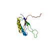
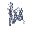
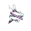
- PDB-2oug: Crystal structure of the RfaH transcription factor at 2.1A resolution -
+ Open data

- Basic information
| Entry | Database: PDB / ID: 2oug | ||||||
|---|---|---|---|---|---|---|---|
| Title | Crystal structure of the RfaH transcription factor at 2.1A resolution | ||||||
 Components Components | Transcriptional activator rfaH | ||||||
 Keywords Keywords | TRANSCRIPTION / transcription factor / virulence / transcription pausing / transcription elongation | ||||||
| Function / homology |  Function and homology information Function and homology informationregulatory RNA binding / transcription antitermination factor activity, DNA binding / translation activator activity / DNA-templated transcription elongation / bacterial-type RNA polymerase core enzyme binding / positive regulation of translation / transcription antitermination / DNA binding / cytosol Similarity search - Function | ||||||
| Biological species |  | ||||||
| Method |  X-RAY DIFFRACTION / SYNCHROTRON / SAD / Resolution: 2.1 Å X-RAY DIFFRACTION / SYNCHROTRON / SAD / Resolution: 2.1 Å | ||||||
 Authors Authors | Vassylyev, D.G. / Vassylyeva, M.N. / Svetlov, V. / Artsimovitch, I. | ||||||
 Citation Citation | Journal: Mol.Cell / Year: 2007 Title: Structural basis for converting a general transcription factor into an operon-specific virulence regulator. Authors: Belogurov, G.A. / Vassylyeva, M.N. / Svetlov, V. / Klyuyev, S. / Grishin, N.V. / Vassylyev, D.G. / Artsimovitch, I. | ||||||
| History |
|
- Structure visualization
| Structure viewer | Molecule: MolmilJmol/JSmol |
|---|
- Downloads & links
-Download
| PDBx/mmCIF format | 2oug.cif.gz | 132.5 KB | Display | PDBx/mmCIF format |
|---|---|---|---|---|
| PDB format | pdb2oug.ent.gz | 105.1 KB | Display | PDB format |
| PDBx/mmJSON format | 2oug.json.gz | Tree view | PDBx/mmJSON format | |
| Others |  Other downloads Other downloads |
-Validation report
| Arichive directory | https://data.pdbj.org/pub/pdb/validation_reports/ou/2ougftp://data.pdbj.org/pub/pdb/validation_reports/ou/2oug | HTTPS FTP |
|---|
-Related structure data
| Similar structure data |
|---|


-Links
 PDBj
PDBj

- Assembly
| Deposited unit | 
| ||||||||
|---|---|---|---|---|---|---|---|---|---|
| 1 | 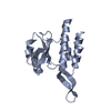
| ||||||||
| 2 | 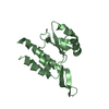
| ||||||||
| 3 | 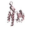
| ||||||||
| 4 | 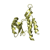
| ||||||||
| Unit cell |
| ||||||||
| Details | The biological assembly is a monomer |
-Components
| #1: Protein | Mass: 18364.217 Da / Num. of mol.: 4 Source method: isolated from a genetically manipulated source Source: (gene. exp.) #2: Water | ChemComp-HOH / |  Mass: 18.015 Da / Num. of mol.: 477 / Source method: isolated from a natural source / Formula: H2O Mass: 18.015 Da / Num. of mol.: 477 / Source method: isolated from a natural source / Formula: H2O |
|---|
-Experimental details
-Experiment
| Experiment | Method: X-RAY DIFFRACTION / Number of used crystals: 1 |
|---|
- Sample preparation
| Crystal | Density Matthews: 2.4 Å3/Da / Density % sol: 48.8 % |
|---|---|
| Crystal grow | Temperature: 277 K / Method: vapor diffusion, sitting drop / pH: 6.5 Details: 20% PEG MME 5000, 50mM sodium cacodylate, 20 mM Co chloride, pH 6.5, VAPOR DIFFUSION, SITTING DROP, temperature 277K |
-Data collection
| Diffraction | Mean temperature: 100 K |
|---|---|
| Diffraction source | Source: SYNCHROTRON / Site: APS  / Beamline: 22-ID / Wavelength: 1 Å / Beamline: 22-ID / Wavelength: 1 Å |
| Detector | Type: MARMOSAIC 300 mm CCD / Detector: CCD / Date: Oct 12, 2005 |
| Radiation | Monochromator: graphite / Protocol: SINGLE WAVELENGTH / Monochromatic (M) / Laue (L): M / Scattering type: x-ray |
| Radiation wavelength | Wavelength: 1 Å / Relative weight: 1 |
| Reflection | Resolution: 2.1→30 Å / Num. all: 38387 / Num. obs: 38387 / % possible obs: 93.8 % / Observed criterion σ(F): 0 / Observed criterion σ(I): 0 / Redundancy: 4.7 % / Biso Wilson estimate: 42 Å2 / Rmerge(I) obs: 0.051 / Rsym value: 0.051 / Net I/σ(I): 27.6 |
| Reflection shell | Resolution: 2.1→2.18 Å / Redundancy: 2.6 % / Rmerge(I) obs: 0.497 / Mean I/σ(I) obs: 2.4 / Num. unique all: 3238 / Rsym value: 0.497 / % possible all: 76.7 |
- Processing
| Software |
| |||||||||||||||||||||||||
|---|---|---|---|---|---|---|---|---|---|---|---|---|---|---|---|---|---|---|---|---|---|---|---|---|---|---|
| Refinement | Method to determine structure: SAD / Resolution: 2.1→30 Å / Isotropic thermal model: isotropic / Cross valid method: THROUGHOUT / σ(F): 0 / σ(I): 0 / Stereochemistry target values: Engh & Huber Details: THE PERFECT MEROHEDRAL TWINNING WAS DETECTED IN THE CRYSTALS WITH THE TWINNING OPERATOR {H,-H,-K,-L}. THE REFINEMENT STATISTICS PRESENTED FOR THIS ENTRY CORRESPONDS TO THE REFINEMENT CARRIED ...Details: THE PERFECT MEROHEDRAL TWINNING WAS DETECTED IN THE CRYSTALS WITH THE TWINNING OPERATOR {H,-H,-K,-L}. THE REFINEMENT STATISTICS PRESENTED FOR THIS ENTRY CORRESPONDS TO THE REFINEMENT CARRIED OUT USING THE TWINNING OPTION OF THE CNS PROGRAM. FOR THE DEPOSITION THE DIFFRACTION DATA WERE DETWINNED USING THE CNS PROGRAM. THEREFORE, SOME REFLECTIONS WERE LOST DUE TO THE DETWINNING PROCEDURE AND ARE MISSING IN THE DEPOSITED SF FILE, WHEREAS THE REFINEMENT STATISTICS CALCULATED BASED ON THE DETWINNED DATA MIGHT BE SLIGHTLY DIFFERENT FROM THOSE OBTAINED DURING THE "TWINNED" REFINEMENT INCLUDED IN THIS ENTRY.
| |||||||||||||||||||||||||
| Displacement parameters | Biso mean: 43.3 Å2 | |||||||||||||||||||||||||
| Refine analyze |
| |||||||||||||||||||||||||
| Refinement step | Cycle: LAST / Resolution: 2.1→30 Å
| |||||||||||||||||||||||||
| Refine LS restraints |
| |||||||||||||||||||||||||
| LS refinement shell | Resolution: 2.1→2.18 Å
|
