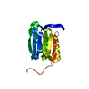
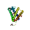
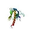
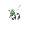
Microtubule-associatedproteins1A/1Blightchain3B / Autophagy-related protein LC3 B / Autophagy-related ubiquitin-like modifier LC3 B / MAP1 light ...Autophagy-related protein LC3 B / Autophagy-related ubiquitin-like modifier LC3 B / MAP1 light chain 3-like protein 2 / MAP1A/MAP1B light chain 3 B / MAP1A/MAP1B LC3 B / Microtubule-associated protein 1 light chain 3 beta
Mass: 15731.887 Da / Num. of mol.: 2 / Fragment: UNP RESIDUES 2-119 Source method: isolated from a genetically manipulated source Source: (gene. exp.) Homo sapiens (human) / Gene: MAP1LC3B, MAP1ALC3 / Plasmid: pET30 / Production host: Escherichia coli (E. coli) / Strain (production host): BL21(DE3) / References: UniProt: Q9GZQ8
Resolution: 2.71→48.8 Å / Cor.coef. Fo:Fc: 0.931 / Cor.coef. Fo:Fc free: 0.893 / SU B: 37.222 / SU ML: 0.352 / Cross valid method: THROUGHOUT / ESU R Free: 0.441 / Stereochemistry target values: MAXIMUM LIKELIHOOD / Details: HYDROGENS HAVE BEEN ADDED IN THE RIDING POSITIONS
Rfactor
Num. reflection
% reflection
Selection details
Rfree
0.29384
354
4.6 %
RANDOM
Rwork
0.22442
-
-
-
obs
0.22752
7343
92.72 %
-
Solvent computation
Ion probe radii: 0.8 Å / Shrinkage radii: 0.8 Å / VDW probe radii: 1.2 Å / Solvent model: MASK
 Movie
Movie Controller


 Open data
 Basic information
 Components
 Keywords
 Function and homology information
 Homo sapiens (human)
 X-RAY DIFFRACTION /
  Authors
 Citation
 Structure visualization
 Downloads & links
 Other downloads
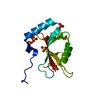


 PDBj
PDBj


 Assembly


 Mass: 18.015 Da / Num. of mol.: 25 / Source method: isolated from a natural source / Formula: H2O
 Sample preparation
 / Beamline: BL-5A / Wavelength: 1 Å
 Processing