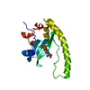
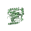
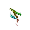
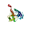
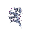
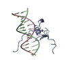
B: DNA (5'-D(*CP*AP*GP*CP*GP*AP*TP*AP*GP*AP*GP*AP*C)-3') C: DNA (5'-D(*GP*TP*CP*TP*CP*TP*AP*TP*CP*GP*CP*TP*G)-3') A: NITROGEN REGULATORY PROTEIN AREA hetero molecules
Mass: 65.409 Da / Num. of mol.: 1 / Source method: obtained synthetically / Formula: Zn
-
Experimental details
-
Experiment
Experiment
Method: SOLUTION NMR
NMR details
Text: DATA WERE RECORDED ON A 1:1 COMPLEX
-
Sample preparation
Sample conditions
pH: 6.1 / Temperature: 298 K
Crystal grow
*PLUS
Method: other / Details: NMR
-
NMR measurement
NMR spectrometer
Type
Manufacturer
Model
Field strength (MHz)
Spectrometer-ID
Bruker AMX500
Bruker
AMX500
360
1
Bruker DMX500
Bruker
DMX500
600
2
Bruker AMX600
Bruker
AMX600
500
3
Bruker DMX600
Bruker
DMX600
750
4
Bruker DMX750
Bruker
DMX750
750
5
Bruker AND AM360
Bruker
ANDAM360
750
6
Bruker 600
Bruker
600
750
7
-
Processing
Software
Name
Version
Classification
X-PLOR
3.1
modelbuilding
X-PLOR
3.1
refinement
X-PLOR
3.1
phasing
NMR software
Name
Version
Developer
Classification
X-PLOR
3.1
BRUNGER
refinement
X-PLOR MODIFIED
MODIFIED
structuresolution
Refinement
Method: simulated annealing / Software ordinal: 1 Details: THE STRUCTURES WERE CALCULATED USING THE SIMULATED ANNEALING PROTOCOL OF NILGES ET AL. (1988) FEBS LETT. 229, 129 - 136 AND PROTEIN ENGINEERING 2, 27 - 38 USING THE PROGRAM X-PLOR MODIFIED ...Details: THE STRUCTURES WERE CALCULATED USING THE SIMULATED ANNEALING PROTOCOL OF NILGES ET AL. (1988) FEBS LETT. 229, 129 - 136 AND PROTEIN ENGINEERING 2, 27 - 38 USING THE PROGRAM X-PLOR MODIFIED TO INCORPORATE COUPLING CONSTANT RESTRAINTS (GARRETT ET AL. (1994) J. MAGN RESON. SERIES B 104, 99 - 103), CARBON CHEMICAL SHIFT RESTRAINTS (KUSZEWSKI ET AL. (1995) J. MAGN. RESON. SERIES B 106, 92 - 96) RESTRAINTS, DIPOLAR COUPLING RESTRAINTS (TJANDRA ET AL. (1997) NATURE STRUCT BIOL 4, 732-738) AND A CONFORMATIONAL DATABASE POTENTIAL FOR PROTEINS AND NUCLEIC ACIDS (KUSZEWSKI ET AL. (1996) PROTEIN SCI 5, 1067 - 1080 AND (1997) J. MAGN. RESON. 125, 171-177) THE 3D STRUCTURE OF THE COMPLEX OF THE WILD TYPE AREA DBD-DNA COMPLEX WAS SOLVED BY MULTI-DIMENSIONAL HETERONUCLEAR-EDITED AND -FILTERED NMR IS BASED ON THE FOLLOWING 1098 EXPERIMENTAL RESTRAINTS (A) PROTEIN: 119 SEQUENTIAL (|I-J|=1), 49 SHORT RANGE (1 < |I-J| >=5), 68 LONG RANGE (|I-J|>5), AND 64 INTRARESIDUE APPROXIMATE INTERPROTON DISTANCE RESTRAINTS; NULL 124 TORSION ANGLE RESTRAINTS (61 PHI, 8 PSI, 39 CHI1, 15 CHI2, AND 1 CHI3), 41 THREE-BOND HN-HA COUPLING CONSTANT RESTRAINTS; NULL 77 (41 CALPHA AND 36 CBETA) 13C CHEMICAL SHIFT RESTRAINTS; NULL 48 RESIDUAL N-H DIPOLAR COUPLING RESTRAINTS; 20 DISTANCE RESTRAINTS FOR 10 BACKBONE HYDROGEN BONDS. (B) DNA: 75 INTRARESIDUE, 115 SEQUENTIAL INTRASTRAND AND 20 INTERSTRAND INTERPROTON DISTANCE RESTRAINTS; 66 DISTANCES FOR WATSON-CRICK BASE PAIR HYDROGEN BONDS; 170 TORSION ANGLE RESTRAINTS FOR THE DNA BACKBONE COVERING VALUES CHARACTERISTIC OF BOTH A AND B DNA. (C) 48 INTERMOLECULAR INTERPROTON DISTANCE RESTRAINTS (D) 2 INTERMOLECULAR DISTANCE RESTRAINTS TO PHOSPHATES (E) 8 'REPULSIVE' RESTRAINTS (F) 4 DISTANCE RESTRAINTS FOR 2 INTERMOLECULAR H-BONDS BETWEEN ARG 24 AND BASE OF GUA5. THE STRUCTURE IN THIS ENTRY IS THE RESTRAINED REGULARIZED MEAN STRUCTURE AND THE LAST NUMERIC COLUMN REPRESENTS THE RMS OF THE 35 INDIVIDUAL SIMULATED ANNEALING STRUCTURES FOUND IN PDB ENTRY 5GAT ABOUT THE MEAN COORDINATE POSITIONS. THE LAST NUMERIC COLUMN IN THE INDIVIDUAL SA STRUCTURES HAS NO MEANING. THE FOLLOWING TWO SETS OF COORDINATES DEFINE THE PRINCIPAL AXIS OF THE MAGNETIC SUSCEPTIBILITY TENSOR: POINT 1 115.014-147.742 -73.887 POINT 2 115.768-146.457 -73.538
NMR ensemble
Conformer selection criteria: REGULARIZED MEAN STRUCTURE / Conformers calculated total number: 35 / Conformers submitted total number: 1
+
About Yorodumi
-
News
-
Feb 9, 2022. New format data for meta-information of EMDB entries
New format data for meta-information of EMDB entries
Version 3 of the EMDB header file is now the official format.
The previous official version 1.9 will be removed from the archive.
In the structure databanks used in Yorodumi, some data are registered as the other names, "COVID-19 virus" and "2019-nCoV". Here are the details of the virus and the list of structure data.
Jan 31, 2019. EMDB accession codes are about to change! (news from PDBe EMDB page)
EMDB accession codes are about to change! (news from PDBe EMDB page)
The allocation of 4 digits for EMDB accession codes will soon come to an end. Whilst these codes will remain in use, new EMDB accession codes will include an additional digit and will expand incrementally as the available range of codes is exhausted. The current 4-digit format prefixed with “EMD-” (i.e. EMD-XXXX) will advance to a 5-digit format (i.e. EMD-XXXXX), and so on. It is currently estimated that the 4-digit codes will be depleted around Spring 2019, at which point the 5-digit format will come into force.
The EM Navigator/Yorodumi systems omit the EMD- prefix.
Related info.:Q: What is EMD? / ID/Accession-code notation in Yorodumi/EM Navigator
Yorodumi is a browser for structure data from EMDB, PDB, SASBDB, etc.
This page is also the successor to EM Navigator detail page, and also detail information page/front-end page for Omokage search.
The word "yorodu" (or yorozu) is an old Japanese word meaning "ten thousand". "mi" (miru) is to see.
Related info.:EMDB / PDB / SASBDB / Comparison of 3 databanks / Yorodumi Search / Aug 31, 2016. New EM Navigator & Yorodumi / Yorodumi Papers / Jmol/JSmol / Function and homology information / Changes in new EM Navigator and Yorodumi
 Movie
Movie Controller

 Yorodumi
 Open data
 Basic information
 Components
 Keywords
 Function and homology information
 Authors
 Citation
 Structure visualization
 Downloads & links
 Other downloads


 PDBj
PDBj


 Assembly


 Mass: 65.409 Da / Num. of mol.: 1 / Source method: obtained synthetically / Formula: Zn
 Sample preparation
 Processing
 X-PLOR