Resolution: 1.8→33.19 Å / Cor.coef. Fo:Fc: 0.96 / Cor.coef. Fo:Fc free: 0.941 / SU B: 5.265 / SU ML: 0.099 / Cross valid method: THROUGHOUT / ESU R: 0.13 / ESU R Free: 0.129 / Stereochemistry target values: MAXIMUM LIKELIHOOD / Details: HYDROGENS HAVE BEEN ADDED IN THE RIDING POSITIONS
Rfactor
Num. reflection
% reflection
Selection details
Rfree
0.23471
1397
5 %
RANDOM
Rwork
0.18913
-
-
-
obs
0.19133
26406
99.91 %
-
Solvent computation
Ion probe radii: 0.8 Å / Shrinkage radii: 0.8 Å / VDW probe radii: 1.2 Å / Solvent model: MASK
Displacement parameters
Biso mean: 36.983 Å2
Baniso -1
Baniso -2
Baniso -3
1-
-0.43 Å2
-0 Å2
0 Å2
2-
-
-0.43 Å2
-0 Å2
3-
-
-
0.86 Å2
Refinement step
Cycle: LAST / Resolution: 1.8→33.19 Å
Protein
Nucleic acid
Ligand
Solvent
Total
Num. atoms
2065
0
78
200
2343
Refine LS restraints
Refine-ID
Type
Dev ideal
Dev ideal target
Number
X-RAY DIFFRACTION
r_bond_refined_d
0.016
0.019
2215
X-RAY DIFFRACTION
r_bond_other_d
0.002
0.02
2145
X-RAY DIFFRACTION
r_angle_refined_deg
1.915
2.02
3038
X-RAY DIFFRACTION
r_angle_other_deg
0.927
3
4944
X-RAY DIFFRACTION
r_dihedral_angle_1_deg
5.908
5
289
X-RAY DIFFRACTION
r_dihedral_angle_2_deg
29.76
24.667
75
X-RAY DIFFRACTION
r_dihedral_angle_3_deg
12.619
15
362
X-RAY DIFFRACTION
r_dihedral_angle_4_deg
14.733
15
10
X-RAY DIFFRACTION
r_chiral_restr
0.101
0.2
376
X-RAY DIFFRACTION
r_gen_planes_refined
0.008
0.021
2409
X-RAY DIFFRACTION
r_gen_planes_other
0.001
0.02
423
X-RAY DIFFRACTION
r_mcbond_it
1.366
1.553
1124
X-RAY DIFFRACTION
r_mcbond_other
1.366
1.553
1123
X-RAY DIFFRACTION
r_mcangle_it
2.263
2.317
1402
X-RAY DIFFRACTION
r_mcangle_other
2.263
2.316
1403
X-RAY DIFFRACTION
r_scbond_it
1.73
1.742
1091
X-RAY DIFFRACTION
r_scbond_other
1.729
1.742
1091
X-RAY DIFFRACTION
r_scangle_other
2.649
2.554
1629
X-RAY DIFFRACTION
r_long_range_B_refined
6.834
14.285
2588
X-RAY DIFFRACTION
r_long_range_B_other
6.833
14.294
2589
LS refinement shell
Resolution: 1.8→1.847 Å / Total num. of bins used: 20
Rfactor
Num. reflection
% reflection
Rfree
0.309
106
-
Rwork
0.264
1897
-
obs
-
-
99.6 %
Refinement TLS params.
Method: refined / Refine-ID: X-RAY DIFFRACTION
ID
L11 (°2)
L12 (°2)
L13 (°2)
L22 (°2)
L23 (°2)
L33 (°2)
S11 (Å °)
S12 (Å °)
S13 (Å °)
S21 (Å °)
S22 (Å °)
S23 (Å °)
S31 (Å °)
S32 (Å °)
S33 (Å °)
T11 (Å2)
T12 (Å2)
T13 (Å2)
T22 (Å2)
T23 (Å2)
T33 (Å2)
Origin x (Å)
Origin y (Å)
Origin z (Å)
1
6.5505
-2.2419
0.8269
3.2828
0.4177
2.3355
0.0346
-0.2284
0.3432
-0.2337
-0.1135
0.3752
-0.0825
-0.3717
0.0789
0.0427
0.0555
-0.0043
0.1802
-0.0055
0.1902
19.078
-39.332
-6.826
2
3.6036
-0.181
-0.7787
5.2995
-0.0204
2.8784
0.162
0.059
-0.2725
-0.159
-0.0988
-0.2799
0.3074
-0.0518
-0.0632
0.0732
0.0132
-0.0018
0.0121
0.0149
0.0596
38.499
-58.8
-11.515
Refinement TLS group
ID
Refine-ID
Refine TLS-ID
Auth asym-ID
Auth seq-ID
1
X-RAY DIFFRACTION
1
A
3 - 144
2
X-RAY DIFFRACTION
2
B
2 - 144
+
About Yorodumi
-
News
-
Feb 9, 2022. New format data for meta-information of EMDB entries
New format data for meta-information of EMDB entries
Version 3 of the EMDB header file is now the official format.
The previous official version 1.9 will be removed from the archive.
In the structure databanks used in Yorodumi, some data are registered as the other names, "COVID-19 virus" and "2019-nCoV". Here are the details of the virus and the list of structure data.
Jan 31, 2019. EMDB accession codes are about to change! (news from PDBe EMDB page)
EMDB accession codes are about to change! (news from PDBe EMDB page)
The allocation of 4 digits for EMDB accession codes will soon come to an end. Whilst these codes will remain in use, new EMDB accession codes will include an additional digit and will expand incrementally as the available range of codes is exhausted. The current 4-digit format prefixed with “EMD-” (i.e. EMD-XXXX) will advance to a 5-digit format (i.e. EMD-XXXXX), and so on. It is currently estimated that the 4-digit codes will be depleted around Spring 2019, at which point the 5-digit format will come into force.
The EM Navigator/Yorodumi systems omit the EMD- prefix.
Related info.:Q: What is EMD? / ID/Accession-code notation in Yorodumi/EM Navigator
Yorodumi is a browser for structure data from EMDB, PDB, SASBDB, etc.
This page is also the successor to EM Navigator detail page, and also detail information page/front-end page for Omokage search.
The word "yorodu" (or yorozu) is an old Japanese word meaning "ten thousand". "mi" (miru) is to see.
Related info.:EMDB / PDB / SASBDB / Comparison of 3 databanks / Yorodumi Search / Aug 31, 2016. New EM Navigator & Yorodumi / Yorodumi Papers / Jmol/JSmol / Function and homology information / Changes in new EM Navigator and Yorodumi
 Movie
Movie Controller

 Yorodumi
 Open data
 Basic information
 Components
 Keywords
 Function and homology information
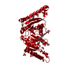
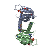
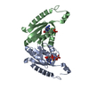
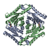
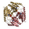
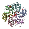
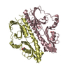
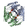
 Salmonella enterica subsp. enterica serovar Typhimurium str. LT2 (bacteria)
 X-RAY DIFFRACTION /
  Authors
 Citation
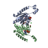
 Structure visualization
 Downloads & links
 Other downloads


 PDBj
PDBj Assembly


 Mass: 24.305 Da / Num. of mol.: 2 / Source method: obtained synthetically / Formula: Mg
 Mass: 507.181 Da / Num. of mol.: 2 / Source method: obtained synthetically / Formula: C10H16N5O13P3 / Comment: ATP, energy-carrying molecule*YM
 Mass: 35.453 Da / Num. of mol.: 2 / Source method: obtained synthetically / Formula: Cl
 Mass: 62.068 Da / Num. of mol.: 3 / Source method: obtained synthetically / Formula: C2H6O2
 Sample preparation
 / Beamline: BM14 / Wavelength: 0.95372 Å
 Processing