 Movie
Movie Controller

[English] 日本語
 Yorodumi
- PDB-1k1e: Structure Of the cobalt-bound form of the deoxy-D-mannose-octulos... -
+ Open data

- Basic information
| Entry | Database: PDB / ID: 1k1e | ||||||
|---|---|---|---|---|---|---|---|
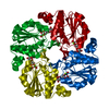
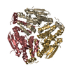
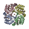
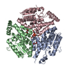
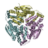
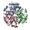
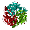
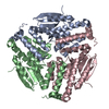
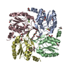
| Title | Structure Of the cobalt-bound form of the deoxy-D-mannose-octulosonate 8-phosphate phosphatase (YrbI) From Haemophilus Influenzae (HI1679) | ||||||
 Components Components | deoxy-D-mannose-octulosonate 8-phosphate phosphatase | ||||||
 Keywords Keywords | HYDROLASE / HI1679 / structural genomics / KDO 8-P phosphatase / Structure 2 Function Project / S2F | ||||||
| Function / homology |  Function and homology information Function and homology information3-deoxy-manno-octulosonate-8-phosphatase / 3-deoxy-manno-octulosonate-8-phosphatase activity / lipopolysaccharide biosynthetic process / metal ion binding Similarity search - Function | ||||||
| Biological species |  Haemophilus influenzae Rd (bacteria) Haemophilus influenzae Rd (bacteria) | ||||||
| Method |  X-RAY DIFFRACTION / SYNCHROTRON / MOLECULAR REPLACEMENT / Resolution: 1.67 Å X-RAY DIFFRACTION / SYNCHROTRON / MOLECULAR REPLACEMENT / Resolution: 1.67 Å | ||||||
 Authors Authors | Lim, K. / Herzberg, O. / Structure 2 Function Project (S2F) | ||||||
 Citation Citation | Journal: Proteins / Year: 2002 Title: From structure to function: YrbI from Haemophilus influenzae (HI1679) is a phosphatase. Authors: Parsons, J.F. / Lim, K. / Tempczyk, A. / Krajewski, W. / Eisenstein, E. / Herzberg, O. | ||||||
| History |
| ||||||
| Remark 400 | COMPOUND THE FUNCTION OF THE PROTEIN WAS ASSIGNED INDEPENDENTLY BY THE WOODARD GROUP: ESCHERICHIA ...COMPOUND THE FUNCTION OF THE PROTEIN WAS ASSIGNED INDEPENDENTLY BY THE WOODARD GROUP: ESCHERICHIA COLI YRBI IS 3-DEOXY-D-MANNO-OCTULOSONATE 8-PHOSPHATE PHOSPHATASE, WU J, WOODARD RW; J BIOL CHEM. 2003 278:18117-2. |
- Structure visualization
| Structure viewer | Molecule: MolmilJmol/JSmol |
|---|
- Downloads & links
-Download
| PDBx/mmCIF format | 1k1e.cif.gz | 437.6 KB | Display | PDBx/mmCIF format |
|---|---|---|---|---|
| PDB format | pdb1k1e.ent.gz | 352.7 KB | Display | PDB format |
| PDBx/mmJSON format | 1k1e.json.gz | Tree view | PDBx/mmJSON format | |
| Others |  Other downloads Other downloads |
-Validation report
| Arichive directory | https://data.pdbj.org/pub/pdb/validation_reports/k1/1k1eftp://data.pdbj.org/pub/pdb/validation_reports/k1/1k1e | HTTPS FTP |
|---|
-Related structure data
| Related structure data |  1j8dSC S: Starting model for refinement C: citing same article ( |
|---|---|
| Similar structure data | |
| Other databases |

-Links
 PDBj
PDBj


- Assembly
| Deposited unit | 
| ||||||||
|---|---|---|---|---|---|---|---|---|---|
| 1 | 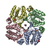
| ||||||||
| 2 | 
| ||||||||
| 3 | 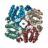
| ||||||||
| 4 | 
| ||||||||
| 5 | 
| ||||||||
| Unit cell |
| ||||||||
| Details | The biological unit is a tetramer. The asymmetric unit contains three tetramers |
-Components
-Protein , 1 types, 12 molecules ABCDEFGHIJKL
| #1: Protein | Mass: 19455.283 Da / Num. of mol.: 12 Source method: isolated from a genetically manipulated source Source: (gene. exp.) Haemophilus influenzae Rd (bacteria) / Species: Haemophilus influenzae / Strain: KW20 / Gene: HI1679 / Plasmid: PDEST17-HI1679 / Production host: References: UniProt: P45314, Hydrolases; Acting on ester bonds; Phosphoric-monoester hydrolases |
|---|
-Non-polymers , 6 types, 1465 molecules 


| #2: Chemical | ChemComp-CO /  Mass: 58.933 Da / Num. of mol.: 12 / Source method: obtained synthetically / Formula: Co Mass: 58.933 Da / Num. of mol.: 12 / Source method: obtained synthetically / Formula: Co#3: Chemical | ChemComp-HG /  Mass: 200.590 Da / Num. of mol.: 12 / Source method: obtained synthetically / Formula: Hg Mass: 200.590 Da / Num. of mol.: 12 / Source method: obtained synthetically / Formula: Hg#4: Chemical | ChemComp-SO4 /  Mass: 96.063 Da / Num. of mol.: 18 / Source method: obtained synthetically / Formula: SO4 Mass: 96.063 Da / Num. of mol.: 18 / Source method: obtained synthetically / Formula: SO4#5: Chemical | ChemComp-GOL /  Mass: 92.094 Da / Num. of mol.: 6 / Source method: obtained synthetically / Formula: C3H8O3 Mass: 92.094 Da / Num. of mol.: 6 / Source method: obtained synthetically / Formula: C3H8O3#6: Chemical |  Mass: 195.237 Da / Num. of mol.: 3 / Source method: obtained synthetically / Formula: C6H13NO4S / Comment: pH buffer*YM Mass: 195.237 Da / Num. of mol.: 3 / Source method: obtained synthetically / Formula: C6H13NO4S / Comment: pH buffer*YM#7: Water | ChemComp-HOH / | Mass: 18.015 Da / Num. of mol.: 1414 / Source method: isolated from a natural source / Formula: H2O |
|---|
-Experimental details
-Experiment
| Experiment | Method: X-RAY DIFFRACTION / Number of used crystals: 1 |
|---|
- Sample preparation
| Crystal | Density Matthews: 2.18 Å3/Da / Density % sol: 43.57 % |
|---|---|
| Crystal grow | Temperature: 295 K / Method: vapor diffusion, hanging drop / pH: 6.5 Details: 1.5 M ammonium sulfate, 0.1 M MES, 10 mM CoCl2. Crystal soaked with 1mM Hg acetate for 3 days. 10% glycerol added for flash-cooling, pH 6.5, VAPOR DIFFUSION, HANGING DROP, temperature 295K |
-Data collection
| Diffraction | Mean temperature: 100 K |
|---|---|
| Diffraction source | Source: SYNCHROTRON / Site: NSLS  / Beamline: X12C / Wavelength: 0.95 Å / Beamline: X12C / Wavelength: 0.95 Å |
| Detector | Type: BRANDEIS / Detector: CCD / Date: Aug 20, 2000 |
| Radiation | Protocol: SINGLE WAVELENGTH / Monochromatic (M) / Laue (L): M / Scattering type: x-ray |
| Radiation wavelength | Wavelength: 0.95 Å / Relative weight: 1 |
| Reflection | Resolution: 1.67→50 Å / Num. all: 199616 / Num. obs: 199616 / % possible obs: 86.1 % / Observed criterion σ(F): 0 / Observed criterion σ(I): 0 / Redundancy: 3.3 % / Biso Wilson estimate: 19 Å2 / Rmerge(I) obs: 0.12 / Net I/σ(I): 11.6 |
| Reflection shell | Resolution: 1.67→1.75 Å / Rmerge(I) obs: 0.213 / % possible all: 49.3 |
- Processing
| Software |
| ||||||||||||||||||||
|---|---|---|---|---|---|---|---|---|---|---|---|---|---|---|---|---|---|---|---|---|---|
| Refinement | Method to determine structure: MOLECULAR REPLACEMENT Starting model: HI1679 (PDB code 1j8d) Resolution: 1.67→20 Å / Cross valid method: THROUGHOUT / σ(F): 2 / Stereochemistry target values: Engh & Huber
| ||||||||||||||||||||
| Displacement parameters | Biso mean: 21 Å2 | ||||||||||||||||||||
| Refinement step | Cycle: LAST / Resolution: 1.67→20 Å
| ||||||||||||||||||||
| Refine LS restraints |
| ||||||||||||||||||||
| LS refinement shell | Resolution: 1.67→1.75 Å / Rfactor Rfree: 0.269 / Rfactor Rwork: 0.206 |
