 Movie
Movie Controller

[English] 日本語
 Yorodumi
- PDB-3e8m: Structure-function Analysis of 2-Keto-3-deoxy-D-glycero-D-galacto... -
+ Open data

- Basic information
| Entry | Database: PDB / ID: 3e8m | ||||||
|---|---|---|---|---|---|---|---|
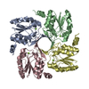
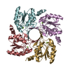
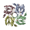
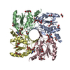
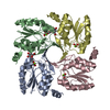
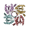
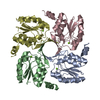
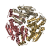
| Title | Structure-function Analysis of 2-Keto-3-deoxy-D-glycero-D-galacto-nononate-9-phosphate (KDN) Phosphatase Defines a New Clad Within the Type C0 HAD Subfamily | ||||||
 Components Components | Acylneuraminate cytidylyltransferase | ||||||
 Keywords Keywords | TRANSFERASE / 2-keto-3-deoxynononic acid 9-phosphate phosphohydrolase / Nucleotidyltransferase | ||||||
| Function / homology |  Function and homology information Function and homology information3-deoxy-D-glycero-D-galacto-nonulopyranosonate 9-phosphatase / hydrolase activity, acting on ester bonds / metal ion binding Similarity search - Function | ||||||
| Biological species |  Bacteroides thetaiotaomicron (bacteria) Bacteroides thetaiotaomicron (bacteria) | ||||||
| Method |  X-RAY DIFFRACTION / SYNCHROTRON / Resolution: 1.1 Å X-RAY DIFFRACTION / SYNCHROTRON / Resolution: 1.1 Å | ||||||
 Authors Authors | Lu, Z. / Wang, L. / Dunaway-Mariano, D. / Allen, K.N. | ||||||
 Citation Citation | Journal: J.Biol.Chem. / Year: 2009 Title: Structure-Function Analysis of 2-Keto-3-deoxy-D-glycero-D-galactonononate-9-phosphate Phosphatase Defines Specificity Elements in Type C0 Haloalkanoate Dehalogenase Family Members. Authors: Lu, Z. / Wang, L. / Dunaway-Mariano, D. / Allen, K.N. | ||||||
| History |
|
- Structure visualization
| Structure viewer | Molecule: MolmilJmol/JSmol |
|---|
- Downloads & links
-Download
| PDBx/mmCIF format | 3e8m.cif.gz | 446.1 KB | Display | PDBx/mmCIF format |
|---|---|---|---|---|
| PDB format | pdb3e8m.ent.gz | 373.8 KB | Display | PDB format |
| PDBx/mmJSON format | 3e8m.json.gz | Tree view | PDBx/mmJSON format | |
| Others |  Other downloads Other downloads |
-Validation report
| Arichive directory | https://data.pdbj.org/pub/pdb/validation_reports/e8/3e8mftp://data.pdbj.org/pub/pdb/validation_reports/e8/3e8m | HTTPS FTP |
|---|
-Related structure data
| Related structure data |  3e81C  3e84C  1k1eS C: citing same article ( S: Starting model for refinement |
|---|---|
| Similar structure data |

-Links
 PDBj
PDBj


- Assembly
| Deposited unit | 
| |||||||||||||||||||||
|---|---|---|---|---|---|---|---|---|---|---|---|---|---|---|---|---|---|---|---|---|---|---|
| 1 |
| |||||||||||||||||||||
| 2 | 
| |||||||||||||||||||||
| Unit cell |
| |||||||||||||||||||||
| Components on special symmetry positions |
|
-Components
-Protein , 1 types, 4 molecules ABCD
| #1: Protein | Mass: 18362.064 Da / Num. of mol.: 4 Source method: isolated from a genetically manipulated source Source: (gene. exp.) Bacteroides thetaiotaomicron (bacteria)Strain: VPI-5482 / Gene: BT_1713 / Plasmid: pET-3A-626 / Production host: |
|---|
-Non-polymers , 8 types, 1119 molecules 


| #2: Chemical | ChemComp-MG /  Mass: 24.305 Da / Num. of mol.: 4 / Source method: obtained synthetically / Formula: Mg Mass: 24.305 Da / Num. of mol.: 4 / Source method: obtained synthetically / Formula: Mg#3: Chemical | ChemComp-CL /  Mass: 35.453 Da / Num. of mol.: 5 / Source method: obtained synthetically / Formula: Cl Mass: 35.453 Da / Num. of mol.: 5 / Source method: obtained synthetically / Formula: Cl#4: Chemical | ChemComp-ACY /  Mass: 60.052 Da / Num. of mol.: 6 / Source method: obtained synthetically / Formula: C2H4O2 Mass: 60.052 Da / Num. of mol.: 6 / Source method: obtained synthetically / Formula: C2H4O2#5: Chemical | ChemComp-PEG /  Mass: 106.120 Da / Num. of mol.: 4 / Source method: obtained synthetically / Formula: C4H10O3 Mass: 106.120 Da / Num. of mol.: 4 / Source method: obtained synthetically / Formula: C4H10O3#6: Chemical | ChemComp-PG4 / |  Mass: 194.226 Da / Num. of mol.: 1 / Source method: obtained synthetically / Formula: C8H18O5 / Comment: precipitant*YM Mass: 194.226 Da / Num. of mol.: 1 / Source method: obtained synthetically / Formula: C8H18O5 / Comment: precipitant*YM#7: Chemical | ChemComp-EDO /  Mass: 62.068 Da / Num. of mol.: 11 / Source method: obtained synthetically / Formula: C2H6O2 Mass: 62.068 Da / Num. of mol.: 11 / Source method: obtained synthetically / Formula: C2H6O2#8: Chemical | ChemComp-PGE / |  Mass: 150.173 Da / Num. of mol.: 1 / Source method: obtained synthetically / Formula: C6H14O4 Mass: 150.173 Da / Num. of mol.: 1 / Source method: obtained synthetically / Formula: C6H14O4#9: Water | ChemComp-HOH / | Mass: 18.015 Da / Num. of mol.: 1087 / Source method: isolated from a natural source / Formula: H2O |
|---|
-Experimental details
-Experiment
| Experiment | Method: X-RAY DIFFRACTION / Number of used crystals: 1 |
|---|
- Sample preparation
| Crystal | Density Matthews: 2.23 Å3/Da / Density % sol: 44.89 % |
|---|---|
| Crystal grow | Temperature: 298 K / Method: vapor diffusion, hanging drop / pH: 8.5 Details: 30% w/v polyethelene glycol (PEG) 4,000, 0.1 M Tris HCl pH 8.5, 0.2 M sodium acetate and 10 mM magnesium chloride, VAPOR DIFFUSION, HANGING DROP, temperature 298.0K |
-Data collection
| Diffraction | Mean temperature: 100 K |
|---|---|
| Diffraction source | Source: SYNCHROTRON / Site: NSLS  / Beamline: X12C / Wavelength: 0.9 Å / Beamline: X12C / Wavelength: 0.9 Å |
| Detector | Type: ADSC QUANTUM 210 / Detector: CCD / Date: Jun 2, 2006 / Details: mirrors |
| Radiation | Monochromator: Si 111 CHANNEL / Protocol: SINGLE WAVELENGTH / Monochromatic (M) / Laue (L): M / Scattering type: x-ray |
| Radiation wavelength | Wavelength: 0.9 Å / Relative weight: 1 |
| Reflection | Resolution: 1.1→50 Å / Num. obs: 258326 / % possible obs: 97.4 % / Observed criterion σ(F): 2 / Observed criterion σ(I): 2 / Redundancy: 11.2 % / Biso Wilson estimate: 8.1 Å2 / Rmerge(I) obs: 0.047 / Χ2: 0.997 / Net I/σ(I): 43.5 |
| Reflection shell | Resolution: 1.1→1.14 Å / Redundancy: 4.2 % / Rmerge(I) obs: 0.29 / Mean I/σ(I) obs: 3.8 / Num. unique all: 22333 / Χ2: 1.011 / % possible all: 84.8 |
- Processing
| Software |
| |||||||||||||||||||||||||||||||||||||||||||||||||
|---|---|---|---|---|---|---|---|---|---|---|---|---|---|---|---|---|---|---|---|---|---|---|---|---|---|---|---|---|---|---|---|---|---|---|---|---|---|---|---|---|---|---|---|---|---|---|---|---|---|---|
| Refinement | Starting model: pdb entry 1K1E Resolution: 1.1→28.782 Å / Occupancy max: 1 / Occupancy min: 0.13 / SU ML: 0.09 / Isotropic thermal model: anisotropic / Cross valid method: FREE R / σ(F): 1.89 / σ(I): 0 / Stereochemistry target values: Engh & Huber
| |||||||||||||||||||||||||||||||||||||||||||||||||
| Solvent computation | Shrinkage radii: 0.9 Å / VDW probe radii: 1.11 Å / Solvent model: FLAT BULK SOLVENT MODEL / Bsol: 47.109 Å2 / ksol: 0.41 e/Å3 | |||||||||||||||||||||||||||||||||||||||||||||||||
| Displacement parameters | Biso max: 102.45 Å2 / Biso mean: 17.685 Å2 / Biso min: 8.24 Å2
| |||||||||||||||||||||||||||||||||||||||||||||||||
| Refine analyze |
| |||||||||||||||||||||||||||||||||||||||||||||||||
| Refinement step | Cycle: LAST / Resolution: 1.1→28.782 Å
| |||||||||||||||||||||||||||||||||||||||||||||||||
| Refine LS restraints |
| |||||||||||||||||||||||||||||||||||||||||||||||||
| LS refinement shell |
|
