 Movie
Movie Controller

[English] 日本語
 Yorodumi
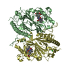
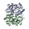
- PDB-4r2k: Crystal structure of H119A mutant of YdaA (Universal Stress Prote... -
+ Open data

- Basic information
| Entry | Database: PDB / ID: 4r2k | ||||||
|---|---|---|---|---|---|---|---|
| Title | Crystal structure of H119A mutant of YdaA (Universal Stress Protein E) from Salmonella typhimurium | ||||||
 Components Components | Universal stress protein E | ||||||
 Keywords Keywords | METAL BINDING PROTEIN / UNKNOWN FUNCTION / Universal stress protein / HUP domain / Internal Symmetry / Stress tolerance / ATP binding / Zinc binding | ||||||
| Function / homology | Rossmann fold - #12370 / UspA / Universal stress protein family / Rossmann fold / 3-Layer(aba) Sandwich / cytoplasm / Alpha Beta / OXALIC ACID / Universal stress protein E Function and homology information Function and homology information | ||||||
| Biological species |  Salmonella enterica subsp. enterica serovar Typhimurium str. LT2 (bacteria) Salmonella enterica subsp. enterica serovar Typhimurium str. LT2 (bacteria) | ||||||
| Method |  X-RAY DIFFRACTION / SYNCHROTRON / MOLECULAR REPLACEMENT / Resolution: 1.97 Å X-RAY DIFFRACTION / SYNCHROTRON / MOLECULAR REPLACEMENT / Resolution: 1.97 Å | ||||||
 Authors Authors | Bangera, M. / Murthy, M.R.N. | ||||||
 Citation Citation | Journal: J.Struct.Biol. / Year: 2015 Title: Structural and functional analysis of two universal stress proteins YdaA and YnaF from Salmonella typhimurium: possible roles in microbial stress tolerance. Authors: Bangera, M. / Panigrahi, R. / Sagurthi, S.R. / Savithri, H.S. / Murthy, M.R. | ||||||
| History |
|
- Structure visualization
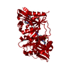
| Structure viewer | Molecule: MolmilJmol/JSmol |
|---|
- Downloads & links
-Download
| PDBx/mmCIF format | 4r2k.cif.gz | 147.8 KB | Display | PDBx/mmCIF format |
|---|---|---|---|---|
| PDB format | pdb4r2k.ent.gz | 115.3 KB | Display | PDB format |
| PDBx/mmJSON format | 4r2k.json.gz | Tree view | PDBx/mmJSON format | |
| Others |  Other downloads Other downloads |
-Validation report
| Arichive directory | https://data.pdbj.org/pub/pdb/validation_reports/r2/4r2kftp://data.pdbj.org/pub/pdb/validation_reports/r2/4r2k | HTTPS FTP |
|---|
-Related structure data
| Related structure data |  4r2jSC 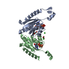 4r2lC 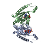 4r2mC S: Starting model for refinement C: citing same article ( |
|---|---|
| Similar structure data |


-Links
 PDBj
PDBj- Assembly
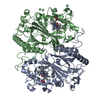
| Deposited unit | 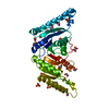
| |||||||||
|---|---|---|---|---|---|---|---|---|---|---|
| 1 | 
| |||||||||
| Unit cell |
| |||||||||
| Components on special symmetry positions |
|
-Components
| #1: Protein | Mass: 37221.477 Da / Num. of mol.: 1 / Mutation: H119A Source method: isolated from a genetically manipulated source Source: (gene. exp.) Salmonella enterica subsp. enterica serovar Typhimurium str. LT2 (bacteria)Strain: LT2 / Gene: STM1661, uspE, ydaA uspE STM1661 / Plasmid: pRSET C / Production host: | ||||||
|---|---|---|---|---|---|---|---|
| #2: Chemical | ChemComp-SO4 /   Mass: 96.063 Da / Num. of mol.: 7 / Source method: obtained synthetically / Formula: SO4 Mass: 96.063 Da / Num. of mol.: 7 / Source method: obtained synthetically / Formula: SO4#3: Chemical | ChemComp-EDO /   Mass: 62.068 Da / Num. of mol.: 16 / Source method: obtained synthetically / Formula: C2H6O2 Mass: 62.068 Da / Num. of mol.: 16 / Source method: obtained synthetically / Formula: C2H6O2#4: Chemical | ChemComp-OXD / | 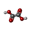  Mass: 90.035 Da / Num. of mol.: 1 / Source method: obtained synthetically / Formula: C2H2O4 Mass: 90.035 Da / Num. of mol.: 1 / Source method: obtained synthetically / Formula: C2H2O4#5: Water | ChemComp-HOH / |  Mass: 18.015 Da / Num. of mol.: 325 / Source method: isolated from a natural source / Formula: H2O Mass: 18.015 Da / Num. of mol.: 325 / Source method: isolated from a natural source / Formula: H2O |
-Experimental details
-Experiment
| Experiment | Method: X-RAY DIFFRACTION / Number of used crystals: 1 |
|---|
- Sample preparation
| Crystal | Density Matthews: 3.25 Å3/Da / Density % sol: 62.13 % |
|---|---|
| Crystal grow | Temperature: 298 K / Method: under oil, microbatch Details: 2M Ammonium sulfate, Under oil, Microbatch, temperature 298.0K |
-Data collection
| Diffraction | Mean temperature: 100 K |
|---|---|
| Diffraction source | Source: SYNCHROTRON / Site: ESRF  / Beamline: BM14 / Wavelength: 0.953725 Å / Beamline: BM14 / Wavelength: 0.953725 Å |
| Detector | Type: MARMOSAIC 225 mm CCD / Detector: CCD / Date: Jul 31, 2012 / Details: bent collimating mirror and toroid |
| Radiation | Monochromator: Si(111) monochromator / Protocol: SINGLE WAVELENGTH / Monochromatic (M) / Laue (L): M / Scattering type: x-ray |
| Radiation wavelength | Wavelength: 0.953725 Å / Relative weight: 1 |
| Reflection | Resolution: 1.97→39.91 Å / Num. obs: 36482 / % possible obs: 100 % / Redundancy: 14.6 % / Rsym value: 0.086 |
| Reflection shell | Resolution: 1.97→2.07 Å / Redundancy: 14.3 % / Mean I/σ(I) obs: 7.3 / Rsym value: 0.348 / % possible all: 100 |
- Processing
| Software |
| ||||||||||||||||||||||||||||||||||||||||||||||||||||||||||||||||||||||||||||||||||||||||||||||||||||||||||||||||||||||||||||||||||||||||||||||||||||||||||||||||||||||||||||||||||||||
|---|---|---|---|---|---|---|---|---|---|---|---|---|---|---|---|---|---|---|---|---|---|---|---|---|---|---|---|---|---|---|---|---|---|---|---|---|---|---|---|---|---|---|---|---|---|---|---|---|---|---|---|---|---|---|---|---|---|---|---|---|---|---|---|---|---|---|---|---|---|---|---|---|---|---|---|---|---|---|---|---|---|---|---|---|---|---|---|---|---|---|---|---|---|---|---|---|---|---|---|---|---|---|---|---|---|---|---|---|---|---|---|---|---|---|---|---|---|---|---|---|---|---|---|---|---|---|---|---|---|---|---|---|---|---|---|---|---|---|---|---|---|---|---|---|---|---|---|---|---|---|---|---|---|---|---|---|---|---|---|---|---|---|---|---|---|---|---|---|---|---|---|---|---|---|---|---|---|---|---|---|---|---|---|
| Refinement | Method to determine structure: MOLECULAR REPLACEMENT Starting model: PDB ID 4R2J Resolution: 1.97→39.46 Å / Cor.coef. Fo:Fc: 0.947 / Cor.coef. Fo:Fc free: 0.927 / SU B: 6.692 / SU ML: 0.103 / Cross valid method: THROUGHOUT / ESU R: 0.133 / ESU R Free: 0.132 / Stereochemistry target values: MAXIMUM LIKELIHOOD / Details: HYDROGENS HAVE BEEN ADDED IN THE RIDING POSITIONS
| ||||||||||||||||||||||||||||||||||||||||||||||||||||||||||||||||||||||||||||||||||||||||||||||||||||||||||||||||||||||||||||||||||||||||||||||||||||||||||||||||||||||||||||||||||||||
| Solvent computation | Ion probe radii: 0.8 Å / Shrinkage radii: 0.8 Å / VDW probe radii: 1.2 Å / Solvent model: MASK | ||||||||||||||||||||||||||||||||||||||||||||||||||||||||||||||||||||||||||||||||||||||||||||||||||||||||||||||||||||||||||||||||||||||||||||||||||||||||||||||||||||||||||||||||||||||
| Displacement parameters | Biso mean: 27.83 Å2
| ||||||||||||||||||||||||||||||||||||||||||||||||||||||||||||||||||||||||||||||||||||||||||||||||||||||||||||||||||||||||||||||||||||||||||||||||||||||||||||||||||||||||||||||||||||||
| Refinement step | Cycle: LAST / Resolution: 1.97→39.46 Å
| ||||||||||||||||||||||||||||||||||||||||||||||||||||||||||||||||||||||||||||||||||||||||||||||||||||||||||||||||||||||||||||||||||||||||||||||||||||||||||||||||||||||||||||||||||||||
| Refine LS restraints |
|