 ムービー
ムービー コントローラー


+ データを開く

- 基本情報
基本情報
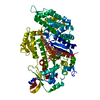
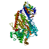
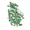
| 登録情報 | データベース: PDB / ID: 6ic3 | ||||||
|---|---|---|---|---|---|---|---|
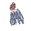
| タイトル | AL amyloid fibril from a lambda 1 light chain | ||||||
 要素 要素 | lambda 1 light chain fragment, residues 3-118 | ||||||
 キーワード キーワード | PROTEIN FIBRIL / amyloid fibril / beta sheet / antibody / heart | ||||||
| 生物種 |  Homo sapiens (ヒト) Homo sapiens (ヒト) | ||||||
| 手法 | 電子顕微鏡法 / らせん対称体再構成法 / クライオ電子顕微鏡法 / 解像度: 3.3 Å | ||||||
 データ登録者 データ登録者 | Fritz, G. / Faendrich, M. / Schmidt, M. / Radamaker, L. | ||||||
 引用 引用 | ジャーナル: Nat Commun / 年: 2019 タイトル: Cryo-EM structure of a light chain-derived amyloid fibril from a patient with systemic AL amyloidosis. 著者: Lynn Radamaker / Yin-Hsi Lin / Karthikeyan Annamalai / Stefanie Huhn / Ute Hegenbart / Stefan O Schönland / Günter Fritz / Matthias Schmidt / Marcus Fändrich /  要旨: Amyloid fibrils derived from antibody light chains are key pathogenic agents in systemic AL amyloidosis. They can be deposited in multiple organs but cardiac amyloid is the major risk factor of ...Amyloid fibrils derived from antibody light chains are key pathogenic agents in systemic AL amyloidosis. They can be deposited in multiple organs but cardiac amyloid is the major risk factor of mortality. Here we report the structure of a λ1 AL amyloid fibril from an explanted human heart at a resolution of 3.3 Å which we determined using cryo-electron microscopy. The fibril core consists of a 91-residue segment presenting an all-beta fold with ten mutagenic changes compared to the germ line. The conformation differs substantially from natively folded light chains: a rotational switch around the intramolecular disulphide bond being the crucial structural rearrangement underlying fibril formation. Our structure provides insight into the mechanism of protein misfolding and the role of patient-specific mutations in pathogenicity. | ||||||
| 履歴 |
|
- 構造の表示
構造の表示
| ムービー |
 ムービービューア ムービービューア |
|---|---|
| 構造ビューア | 分子: MolmilJmol/JSmol |
- ダウンロードとリンク
-ダウンロード
| PDBx/mmCIF形式 | 6ic3.cif.gz | 128.9 KB | 表示 | PDBx/mmCIF形式 |
|---|---|---|---|---|
| PDB形式 | pdb6ic3.ent.gz | 101.6 KB | 表示 | PDB形式 |
| PDBx/mmJSON形式 | 6ic3.json.gz | ツリー表示 | PDBx/mmJSON形式 | |
| その他 |  その他のダウンロード その他のダウンロード |
-検証レポート
| 文書・要旨 | 6ic3_validation.pdf.gz | 724.6 KB | 表示 | wwPDB検証レポート |
|---|---|---|---|---|
| 文書・詳細版 | 6ic3_full_validation.pdf.gz | 732.8 KB | 表示 | |
| XML形式データ | 6ic3_validation.xml.gz | 27.6 KB | 表示 | |
| CIF形式データ | 6ic3_validation.cif.gz | 42 KB | 表示 | |
| アーカイブディレクトリ | https://data.pdbj.org/pub/pdb/validation_reports/ic/6ic3ftp://data.pdbj.org/pub/pdb/validation_reports/ic/6ic3 | HTTPS FTP |
-関連構造データ
| 関連構造データ | 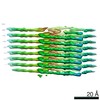 4452MC M: このデータのモデリングに利用したマップデータ C: 同じ文献を引用 ( |
|---|---|
| 類似構造データ | |
| 電子顕微鏡画像生データ | EMPIAR-10245 (タイトル: AL amyloid fibril from a lambda 1 light chain / Data size: 44.7 Data #1: Aligned, motion-corrected micrographs of AL fibrils extracted from human heart tissue [micrographs - single frame]) |


-リンク
 PDBj
PDBj


- 集合体
集合体
| 登録構造単位 | 
|
|---|---|
| 1 |
|
-要素
| #1: 抗体 | 分子量: 12177.463 Da / 分子数: 8 / 由来タイプ: 天然 / 由来: (天然) Homo sapiens (ヒト) / 器官: Heart / 組織: heart muscleHas protein modification | Y | |
|---|
-実験情報
-実験
| 実験 | 手法: 電子顕微鏡法 |
|---|---|
| EM実験 | 試料の集合状態: FILAMENT / 3次元再構成法: らせん対称体再構成法 |
- 試料調製
試料調製
| 構成要素 | 名称: Amyloid fibril of an antibody lambda 1 light chain / タイプ: COMPLEX 詳細: Extracted fibrils from the heart of a patient suffering from systemic AL amyloidosis Entity ID: all / 由来: NATURAL |
|---|---|
| 分子量 | 値: 2.5 kDa/nm / 実験値: YES |
| 由来(天然) | 生物種: Homo sapiens (ヒト) / 器官: heart / 組織: heart muscle |
| 緩衝液 | pH: 7 |
| 緩衝液成分 | 名称: distilled water / 式: H2O |
| 試料 | 包埋: NO / シャドウイング: NO / 染色: NO / 凍結: YES / 詳細: Sample in pure water, pH not determined |
| 試料支持 | グリッドの材料: COPPER / グリッドのサイズ: 400 divisions/in. / グリッドのタイプ: C-flat-1.2/1.3 4C |
| 急速凍結 | 装置: GATAN CRYOPLUNGE 3 / 凍結剤: ETHANE / 湿度: 80 % / 凍結前の試料温度: 294 K / 詳細: blotted from the backside for 4 s before plunging |
- 電子顕微鏡撮影
| 実験機器 |  モデル: Titan Krios / 画像提供: FEI Company |
|---|---|
| 顕微鏡 | モデル: FEI TITAN KRIOS |
| 電子銃 | 電子線源:  FIELD EMISSION GUN / 加速電圧: 300 kV / 照射モード: FLOOD BEAM FIELD EMISSION GUN / 加速電圧: 300 kV / 照射モード: FLOOD BEAM |
| 電子レンズ | モード: BRIGHT FIELD / Cs: 2.7 mm |
| 試料ホルダ | 凍結剤: NITROGEN 試料ホルダーモデル: FEI TITAN KRIOS AUTOGRID HOLDER |
| 撮影 | 平均露光時間: 6 sec. / 電子線照射量: 32 e/Å2 / 検出モード: COUNTING フィルム・検出器のモデル: GATAN K2 SUMMIT (4k x 4k) 撮影したグリッド数: 1 / 実像数: 847 |
| 画像スキャン | 横: 3838 / 縦: 3710 / 動画フレーム数/画像: 30 / 利用したフレーム数/画像: 1-30 |
- 解析
解析
| ソフトウェア | 名称: PHENIX / バージョン: 1.14_3260: / 分類: 精密化 | |||||||||||||||||||||||||||||||||||||||||||||
|---|---|---|---|---|---|---|---|---|---|---|---|---|---|---|---|---|---|---|---|---|---|---|---|---|---|---|---|---|---|---|---|---|---|---|---|---|---|---|---|---|---|---|---|---|---|---|
| EMソフトウェア |
| |||||||||||||||||||||||||||||||||||||||||||||
| 画像処理 | 詳細: Motion-corrected and dose-weighted movie frames | |||||||||||||||||||||||||||||||||||||||||||||
| CTF補正 | 詳細: CTF was estimated from the non-dose-weighted micrographs タイプ: PHASE FLIPPING AND AMPLITUDE CORRECTION | |||||||||||||||||||||||||||||||||||||||||||||
| らせん対称 | 回転角度/サブユニット: 0.58 ° / 軸方向距離/サブユニット: 4.81 Å / らせん対称軸の対称性: C1 | |||||||||||||||||||||||||||||||||||||||||||||
| 粒子像の選択 | 選択した粒子像数: 119395 詳細: manual selection. Sigma contrast 3, lowpass filter 20 A | |||||||||||||||||||||||||||||||||||||||||||||
| 3次元再構成 | 解像度: 3.3 Å / 解像度の算出法: FSC 0.143 CUT-OFF / 粒子像の数: 32677 / アルゴリズム: FOURIER SPACE / クラス平均像の数: 1 / 対称性のタイプ: HELICAL | |||||||||||||||||||||||||||||||||||||||||||||
| 原子モデル構築 | B value: 111.4 / プロトコル: OTHER / 空間: REAL Target criteria: REAL-SPACE (WEIGHTED MAP SUM AT ATOM CENTERS) 詳細: Refinement strategy included global minimization and local grid search and ADP were refined. Secondary structure restraints and NCS were applied during refinement. |
