 Movie
Movie Controller


+ Open data

- Basic information
| Entry | Database: PDB / ID: 6i9z | ||||||
|---|---|---|---|---|---|---|---|
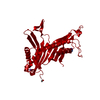
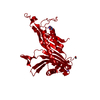
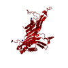
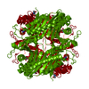
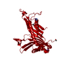
| Title | urate oxidase under 65 bar of argon | ||||||
 Components Components | Uricase | ||||||
 Keywords Keywords | OXIDOREDUCTASE / aspergillus flavus / homotetramer / purine metabolism / argon / high pressure | ||||||
| Function / homology |  Function and homology information Function and homology informationfactor-independent urate hydroxylase / urate oxidase activity / purine nucleobase catabolic process / urate catabolic process / peroxisome Similarity search - Function | ||||||
| Biological species |  | ||||||
| Method |  X-RAY DIFFRACTION / SYNCHROTRON / FOURIER SYNTHESIS / Resolution: 1.6 Å X-RAY DIFFRACTION / SYNCHROTRON / FOURIER SYNTHESIS / Resolution: 1.6 Å | ||||||
 Authors Authors | Prange, T. / Colloc'h, N. / Carpentier, P. | ||||||
 Citation Citation | Journal: Acta Crystallogr D Struct Biol / Year: 2022 Title: Comparative study of the effects of high hydrostatic pressure per se and high argon pressure on urate oxidase ligand stabilization. Authors: Prange, T. / Carpentier, P. / Dhaussy, A.C. / van der Linden, P. / Girard, E. / Colloc'h, N. #1: Journal: Current trends in X-ray crystallography / Year: 2011Title: Protein-noble gas interactions investigated by crystallography on three enzymes - Implication on anesthesia and neuroprotection mechanism. Authors: Colloc'h, N. / Marassio, G. / Prange, T. #2: Journal: J. Applied Crystallography / Year: 2016Title: Gas-sensitive biological crystals processed in pressurized oxygen and krypton atmospheres: deciphering gas channels in proteins using a novel 'soak-and-freeze' methodology Authors: Lafumat, B. / Mueller-Dieckmann, C. / Leonard, G. / Colloc'h, N. / Prange, T. / Giraud, T. / Dobbias, F. / Royant, R. / van der Linden, P. / Carpentier, P. | ||||||
| History |
|
- Structure visualization
| Structure viewer | Molecule: MolmilJmol/JSmol |
|---|
- Downloads & links
-Download
| PDBx/mmCIF format | 6i9z.cif.gz | 81.3 KB | Display | PDBx/mmCIF format |
|---|---|---|---|---|
| PDB format | pdb6i9z.ent.gz | 59 KB | Display | PDB format |
| PDBx/mmJSON format | 6i9z.json.gz | Tree view | PDBx/mmJSON format | |
| Others |  Other downloads Other downloads |
-Validation report
| Arichive directory | https://data.pdbj.org/pub/pdb/validation_reports/i9/6i9zftp://data.pdbj.org/pub/pdb/validation_reports/i9/6i9z | HTTPS FTP |
|---|
-Related structure data
| Related structure data |  6i9xC 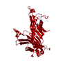 6ia1C 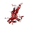 6ia3C 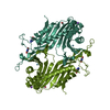 6ia9C  7p0cC  7p0dC  7p0gC 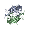 7pufC  7pwnC 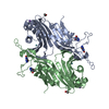 7q09C  1r51S S: Starting model for refinement C: citing same article ( |
|---|---|
| Similar structure data |


-Links
 PDBj
PDBj
- Assembly
| Deposited unit | 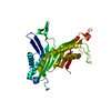
| ||||||||
|---|---|---|---|---|---|---|---|---|---|
| 1 | 
| ||||||||
| Unit cell |
|
-Components
| #1: Protein | Mass: 34183.590 Da / Num. of mol.: 1 Source method: isolated from a genetically manipulated source Details: 6 RESIDUES AT THE C-END NOT OBSERVED IN THE DENSITY: SER-LEU-LYS-SER-LYS-LEU 296- ..... ...-301 Source: (gene. exp.)  References: UniProt: Q00511, factor-independent urate hydroxylase |
|---|---|
| #2: Chemical | ChemComp-AZA /   Mass: 153.099 Da / Num. of mol.: 1 / Source method: obtained synthetically / Formula: C4H3N5O2 Mass: 153.099 Da / Num. of mol.: 1 / Source method: obtained synthetically / Formula: C4H3N5O2 |
| #3: Chemical | ChemComp-NA /   Mass: 22.990 Da / Num. of mol.: 1 / Source method: obtained synthetically / Formula: Na Mass: 22.990 Da / Num. of mol.: 1 / Source method: obtained synthetically / Formula: Na |
| #4: Chemical | ChemComp-AR /   Mass: 39.948 Da / Num. of mol.: 1 / Source method: obtained synthetically / Formula: Ar Mass: 39.948 Da / Num. of mol.: 1 / Source method: obtained synthetically / Formula: Ar |
| #5: Water | ChemComp-HOH /  Mass: 18.015 Da / Num. of mol.: 221 / Source method: isolated from a natural source / Formula: H2O Mass: 18.015 Da / Num. of mol.: 221 / Source method: isolated from a natural source / Formula: H2O |
| Has protein modification | Y |
-Experimental details
-Experiment
| Experiment | Method: X-RAY DIFFRACTION / Number of used crystals: 1 |
|---|
- Sample preparation
| Crystal | Density Matthews: 2.9 Å3/Da / Density % sol: 57.5 % / Description: LARGE COLORLESS PRISMS |
|---|---|
| Crystal grow | Temperature: 291 K / Method: batch mode / pH: 8 Details: 20 microliter of protein (15 mg/ml) mixed with 20 microliter of solution: Tris 0.05M (chloride free) + 4% PEG 4000. |
-Data collection
| Diffraction | Mean temperature: 291 K / Serial crystal experiment: N |
|---|---|
| Diffraction source | Source: SYNCHROTRON / Site: MAX II  / Beamline: I711 / Wavelength: 0.9775 Å / Beamline: I711 / Wavelength: 0.9775 Å |
| Detector | Type: MARMOSAIC 225 mm CCD / Detector: CCD / Date: Jul 14, 2005 |
| Radiation | Monochromator: Si(111) / Protocol: SINGLE WAVELENGTH / Monochromatic (M) / Laue (L): M / Scattering type: x-ray |
| Radiation wavelength | Wavelength: 0.9775 Å / Relative weight: 1 |
| Reflection | Resolution: 1.6→20 Å / Num. obs: 52954 / % possible obs: 98 % / Redundancy: 3.5 % / Rmerge(I) obs: 0.053 / Net I/σ(I): 19.3 |
| Reflection shell | Resolution: 1.6→1.66 Å / Redundancy: 3.3 % / Rmerge(I) obs: 0.5 / Mean I/σ(I) obs: 2.2 / Num. unique obs: 5301 / % possible all: 99.5 |
- Processing
| Software |
| ||||||||||||||||||||||||||||||||||||||||||||||||||||||||||||||||||||||||||||||||||||||||||||||||||||||||||||||||||||||||||||||||||||||||||||||||||||||||||||||||||||||||||||||||||||||
|---|---|---|---|---|---|---|---|---|---|---|---|---|---|---|---|---|---|---|---|---|---|---|---|---|---|---|---|---|---|---|---|---|---|---|---|---|---|---|---|---|---|---|---|---|---|---|---|---|---|---|---|---|---|---|---|---|---|---|---|---|---|---|---|---|---|---|---|---|---|---|---|---|---|---|---|---|---|---|---|---|---|---|---|---|---|---|---|---|---|---|---|---|---|---|---|---|---|---|---|---|---|---|---|---|---|---|---|---|---|---|---|---|---|---|---|---|---|---|---|---|---|---|---|---|---|---|---|---|---|---|---|---|---|---|---|---|---|---|---|---|---|---|---|---|---|---|---|---|---|---|---|---|---|---|---|---|---|---|---|---|---|---|---|---|---|---|---|---|---|---|---|---|---|---|---|---|---|---|---|---|---|---|---|
| Refinement | Method to determine structure: FOURIER SYNTHESIS Starting model: 1R51 Resolution: 1.6→14.32 Å / Cor.coef. Fo:Fc: 0.98 / Cor.coef. Fo:Fc free: 0.97 / SU B: 1.673 / SU ML: 0.055 / Cross valid method: THROUGHOUT / ESU R: 0.066 / ESU R Free: 0.069 / Details: HYDROGENS HAVE BEEN ADDED IN THE RIDING POSITIONS
| ||||||||||||||||||||||||||||||||||||||||||||||||||||||||||||||||||||||||||||||||||||||||||||||||||||||||||||||||||||||||||||||||||||||||||||||||||||||||||||||||||||||||||||||||||||||
| Solvent computation | Ion probe radii: 0.8 Å / Shrinkage radii: 0.8 Å / VDW probe radii: 1.2 Å | ||||||||||||||||||||||||||||||||||||||||||||||||||||||||||||||||||||||||||||||||||||||||||||||||||||||||||||||||||||||||||||||||||||||||||||||||||||||||||||||||||||||||||||||||||||||
| Displacement parameters | Biso mean: 24.744 Å2
| ||||||||||||||||||||||||||||||||||||||||||||||||||||||||||||||||||||||||||||||||||||||||||||||||||||||||||||||||||||||||||||||||||||||||||||||||||||||||||||||||||||||||||||||||||||||
| Refinement step | Cycle: 1 / Resolution: 1.6→14.32 Å
| ||||||||||||||||||||||||||||||||||||||||||||||||||||||||||||||||||||||||||||||||||||||||||||||||||||||||||||||||||||||||||||||||||||||||||||||||||||||||||||||||||||||||||||||||||||||
| Refine LS restraints |
|