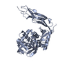
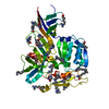
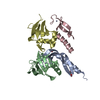
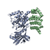
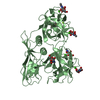
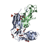
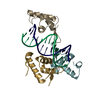
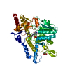
Journal: Nat Commun / Year: 2019 Title: Cryo-EM structure of cardiac amyloid fibrils from an immunoglobulin light chain AL amyloidosis patient. Authors: Paolo Swuec / Francesca Lavatelli / Masayoshi Tasaki / Cristina Paissoni / Paola Rognoni / Martina Maritan / Francesca Brambilla / Paolo Milani / Pierluigi Mauri / Carlo Camilloni / Giovanni ...Authors: Paolo Swuec / Francesca Lavatelli / Masayoshi Tasaki / Cristina Paissoni / Paola Rognoni / Martina Maritan / Francesca Brambilla / Paolo Milani / Pierluigi Mauri / Carlo Camilloni / Giovanni Palladini / Giampaolo Merlini / Stefano Ricagno / Martino Bolognesi / Abstract: Systemic light chain amyloidosis (AL) is a life-threatening disease caused by aggregation and deposition of monoclonal immunoglobulin light chains (LC) in target organs. Severity of heart ...Systemic light chain amyloidosis (AL) is a life-threatening disease caused by aggregation and deposition of monoclonal immunoglobulin light chains (LC) in target organs. Severity of heart involvement is the most important factor determining prognosis. Here, we report the 4.0 Å resolution cryo-electron microscopy map and molecular model of amyloid fibrils extracted from the heart of an AL amyloidosis patient with severe amyloid cardiomyopathy. The helical fibrils are composed of a single protofilament, showing typical 4.9 Å stacking and cross-β architecture. Two distinct polypeptide stretches (total of 77 residues) from the LC variable domain (V) fit the fibril density. Despite V high sequence variability, residues stabilizing the fibril core are conserved through different cardiotoxic V, highlighting structural motifs that may be common to misfolding-prone LCs. Our data shed light on the architecture of LC amyloids, correlate amino acid sequences with fibril assembly, providing the grounds for development of innovative medicines.
Instrument: FEI VITROBOT MARK IV / Cryogen name: ETHANE / Humidity: 100 % / Chamber temperature: 278 K
-
Electron microscopy imaging
Experimental equipment
Model: Talos Arctica / Image courtesy: FEI Company
Microscopy
Model: FEI TALOS ARCTICA
Electron gun
Electron source: FIELD EMISSION GUN / Accelerating voltage: 200 kV / Illumination mode: SPOT SCAN
Electron lens
Mode: BRIGHT FIELD / Nominal magnification: 120000 X / Cs: 2.7 mm / C2 aperture diameter: 50 µm
Specimen holder
Cryogen: NITROGEN / Specimen holder model: OTHER
Image recording
Average exposure time: 1 sec. / Electron dose: 50 e/Å2 / Detector mode: INTEGRATING / Film or detector model: FEI FALCON III (4k x 4k) / Num. of real images: 1680
In the structure databanks used in Yorodumi, some data are registered as the other names, "COVID-19 virus" and "2019-nCoV". Here are the details of the virus and the list of structure data.
Jan 31, 2019. EMDB accession codes are about to change! (news from PDBe EMDB page)
EMDB accession codes are about to change! (news from PDBe EMDB page)
The allocation of 4 digits for EMDB accession codes will soon come to an end. Whilst these codes will remain in use, new EMDB accession codes will include an additional digit and will expand incrementally as the available range of codes is exhausted. The current 4-digit format prefixed with “EMD-” (i.e. EMD-XXXX) will advance to a 5-digit format (i.e. EMD-XXXXX), and so on. It is currently estimated that the 4-digit codes will be depleted around Spring 2019, at which point the 5-digit format will come into force.
The EM Navigator/Yorodumi systems omit the EMD- prefix.
Related info.:Q: What is EMD? / ID/Accession-code notation in Yorodumi/EM Navigator
Yorodumi is a browser for structure data from EMDB, PDB, SASBDB, etc.
This page is also the successor to EM Navigator detail page, and also detail information page/front-end page for Omokage search.
The word "yorodu" (or yorozu) is an old Japanese word meaning "ten thousand". "mi" (miru) is to see.
Related info.:EMDB / PDB / SASBDB / Comparison of 3 databanks / Yorodumi Search / Aug 31, 2016. New EM Navigator & Yorodumi / Yorodumi Papers / Jmol/JSmol / Function and homology information / Changes in new EM Navigator and Yorodumi
 Movie
Movie Controller

 Yorodumi
 Open data
 Basic information
 Components
 Keywords
 Homo sapiens (human)
 Authors
 Italy, 4items
  Citation
 Structure visualization
 Movie viewer
 Downloads & links
 Other downloads
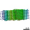


 PDBj
PDBj


 Assembly
 Sample preparation
 Electron microscopy imaging
 FIELD EMISSION GUN / Accelerating voltage: 200 kV / Illumination mode: SPOT SCAN
 Processing