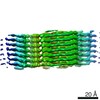
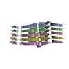
Journal: J Mol Biol / Year: 2023 Title: The Cryo-EM STRUCTURE of Renal Amyloid Fibril Suggests Structurally Homogeneous Multiorgan Aggregation in AL Amyloidosis. Authors: Sarita Puri / Tim Schulte / Antonio Chaves-Sanjuan / Giulia Mazzini / Serena Caminito / Carlo Pappone / Luigi Anastasia / Paolo Milani / Giampaolo Merlini / Martino Bolognesi / Mario ...Authors: Sarita Puri / Tim Schulte / Antonio Chaves-Sanjuan / Giulia Mazzini / Serena Caminito / Carlo Pappone / Luigi Anastasia / Paolo Milani / Giampaolo Merlini / Martino Bolognesi / Mario Nuvolone / Giovanni Palladini / Stefano Ricagno / Abstract: Immunoglobulin light chain amyloidosis (AL) is caused by the aberrant production of amyloidogenic light chains (LC) that accumulate as amyloid deposits in vital organs. Distinct LC sequences in each ...Immunoglobulin light chain amyloidosis (AL) is caused by the aberrant production of amyloidogenic light chains (LC) that accumulate as amyloid deposits in vital organs. Distinct LC sequences in each patient yield distinct amyloid structures. However different tissue microenvironments may also cause identical protein precursors to adopt distinct amyloid structures. To address the impact of the tissue environment on the structural polymorphism of amyloids, we extracted fibrils from the kidney of an AL patient (AL55) whose cardiac amyloid structure was previously determined by our group. Here we show that the 4.0 Å resolution cryo-EM structure of the renal fibril is virtually identical to that reported for the cardiac fibril. These results provide the first structural evidence that LC amyloids independently deposited in different organs of the same AL patient share a common fold.
Mass: 23326.557 Da / Num. of mol.: 5 / Mutation: No / Source method: isolated from a natural source / Source: (natural) Homo sapiens (human) / Organ: Kidney
Has protein modification
Y
-
Experimental details
-
Experiment
Experiment
Method: ELECTRON MICROSCOPY
EM experiment
Aggregation state: FILAMENT / 3D reconstruction method: helical reconstruction
-
Sample preparation
Component
Name: Renal AL55 amyloid fibrils / Type: COMPLEX Details: Light chain amyloid fibrils named AL55 extracted from the kidney of an AL amyloidosis patient Entity ID: all / Source: NATURAL
Molecular weight
ID
Entity assembly-ID
Experimental value
1
1
NO
2
1
NO
Source (natural)
Organism: Homo sapiens (human) / Organ: Kidney
Buffer solution
pH: 7
Specimen
Embedding applied: NO / Shadowing applied: NO / Staining applied: NO / Vitrification applied: YES
In the structure databanks used in Yorodumi, some data are registered as the other names, "COVID-19 virus" and "2019-nCoV". Here are the details of the virus and the list of structure data.
Jan 31, 2019. EMDB accession codes are about to change! (news from PDBe EMDB page)
EMDB accession codes are about to change! (news from PDBe EMDB page)
The allocation of 4 digits for EMDB accession codes will soon come to an end. Whilst these codes will remain in use, new EMDB accession codes will include an additional digit and will expand incrementally as the available range of codes is exhausted. The current 4-digit format prefixed with “EMD-” (i.e. EMD-XXXX) will advance to a 5-digit format (i.e. EMD-XXXXX), and so on. It is currently estimated that the 4-digit codes will be depleted around Spring 2019, at which point the 5-digit format will come into force.
The EM Navigator/Yorodumi systems omit the EMD- prefix.
Related info.:Q: What is EMD? / ID/Accession-code notation in Yorodumi/EM Navigator
Yorodumi is a browser for structure data from EMDB, PDB, SASBDB, etc.
This page is also the successor to EM Navigator detail page, and also detail information page/front-end page for Omokage search.
The word "yorodu" (or yorozu) is an old Japanese word meaning "ten thousand". "mi" (miru) is to see.
Related info.:EMDB / PDB / SASBDB / Comparison of 3 databanks / Yorodumi Search / Aug 31, 2016. New EM Navigator & Yorodumi / Yorodumi Papers / Jmol/JSmol / Function and homology information / Changes in new EM Navigator and Yorodumi
 Movie
Movie Controller

 Yorodumi
 Open data
 Basic information
 Components
 Keywords
 Homo sapiens (human)
 Authors
 Italy, 1items
  Citation
 Structure visualization
 Molmil
 Downloads & links
 Other downloads
 PDBj
PDBj

 Assembly
 Sample preparation
 Electron microscopy imaging
 FIELD EMISSION GUN / Accelerating voltage: 200 kV / Illumination mode: FLOOD BEAM
 Processing