 Movie
Movie Controller

[English] 日本語
 Yorodumi
- PDB-6gn0: Exoenzyme S from Pseudomonas aeruginosa in complex with human 14-... -
+ Open data

- Basic information
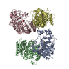
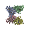
| Entry | Database: PDB / ID: 6gn0 | ||||||
|---|---|---|---|---|---|---|---|
| Title | Exoenzyme S from Pseudomonas aeruginosa in complex with human 14-3-3 protein beta, tetrameric crystal form | ||||||
 Components Components |
| ||||||
 Keywords Keywords | TOXIN / EXOS / PSEUDOMONAS AERUGINOSA / ADP-RIBOSYLATION / NAD | ||||||
| Function / homology |  Function and homology information Function and homology informationnegative regulation of cell growth involved in contact inhibition / Tristetraprolin (TTP, ZFP36) binds and destabilizes mRNA / positive regulation of hippo signaling / Butyrate Response Factor 1 (BRF1) binds and destabilizes mRNA / MTOR signalling / ARMS-mediated activation / SHOC2 M1731 mutant abolishes MRAS complex function / Gain-of-function MRAS complexes activate RAF signaling / Rap1 signalling / Signaling by Hippo ...negative regulation of cell growth involved in contact inhibition / Tristetraprolin (TTP, ZFP36) binds and destabilizes mRNA / positive regulation of hippo signaling / Butyrate Response Factor 1 (BRF1) binds and destabilizes mRNA / MTOR signalling / ARMS-mediated activation / SHOC2 M1731 mutant abolishes MRAS complex function / Gain-of-function MRAS complexes activate RAF signaling / Rap1 signalling / Signaling by Hippo / vacuolar membrane / negative regulation of G protein-coupled receptor signaling pathway / negative regulation of protein import into nucleus / Frs2-mediated activation / protein phosphatase inhibitor activity / glycosyltransferase activity / protein kinase inhibitor activity / Regulation of localization of FOXO transcription factors / mTORC1-mediated signalling / Activation of BAD and translocation to mitochondria / phosphoserine residue binding / SARS-CoV-2 targets host intracellular signalling and regulatory pathways / SARS-CoV-1 targets host intracellular signalling and regulatory pathways / RHO GTPases activate PKNs / protein targeting / Chk1/Chk2(Cds1) mediated inactivation of Cyclin B:Cdk1 complex / transcription repressor complex / nucleotidyltransferase activity / Transcriptional and post-translational regulation of MITF-M expression and activity / GTPase activator activity / TP53 Regulates Metabolic Genes / Translocation of SLC2A4 (GLUT4) to the plasma membrane / protein sequestering activity / phosphoprotein binding / RAF activation / Signaling by high-kinase activity BRAF mutants / MAP2K and MAPK activation / histone deacetylase binding / Signaling by RAF1 mutants / Signaling by moderate kinase activity BRAF mutants / Paradoxical activation of RAF signaling by kinase inactive BRAF / Signaling downstream of RAS mutants / Negative regulation of MAPK pathway / Signaling by BRAF and RAF1 fusions / melanosome / intracellular protein localization / toxin activity / cadherin binding / protein domain specific binding / focal adhesion / negative regulation of DNA-templated transcription / protein-containing complex binding / perinuclear region of cytoplasm / enzyme binding / signal transduction / positive regulation of transcription by RNA polymerase II / extracellular exosome / extracellular region / membrane / identical protein binding / nucleus / cytoplasm / cytosol Similarity search - Function | ||||||
| Biological species |  Homo sapiens (human) Homo sapiens (human)  Pseudomonas aeruginosa (bacteria) Pseudomonas aeruginosa (bacteria) | ||||||
| Method |  X-RAY DIFFRACTION / SYNCHROTRON / MOLECULAR REPLACEMENT / Resolution: 3.24 Å X-RAY DIFFRACTION / SYNCHROTRON / MOLECULAR REPLACEMENT / Resolution: 3.24 Å | ||||||
 Authors Authors | Karlberg, T. / Pinto, A.F. / Hornyak, P. / Nareoja, K. / Schuler, H. | ||||||
| Funding support |  Sweden, 1items Sweden, 1items
| ||||||
 Citation Citation | Journal: Nat Commun / Year: 2018 Title: 14-3-3 proteins activate Pseudomonas exotoxins-S and -T by chaperoning a hydrophobic surface. Authors: Karlberg, T. / Hornyak, P. / Pinto, A.F. / Milanova, S. / Ebrahimi, M. / Lindberg, M. / Pullen, N. / Nordstrom, A. / Loverli, E. / Caraballo, R. / Wong, E.V. / Nareoja, K. / Thorsell, A.G. / ...Authors: Karlberg, T. / Hornyak, P. / Pinto, A.F. / Milanova, S. / Ebrahimi, M. / Lindberg, M. / Pullen, N. / Nordstrom, A. / Loverli, E. / Caraballo, R. / Wong, E.V. / Nareoja, K. / Thorsell, A.G. / Elofsson, M. / De La Cruz, E.M. / Bjorkegren, C. / Schuler, H. | ||||||
| History |
|
- Structure visualization
| Structure viewer | Molecule: MolmilJmol/JSmol |
|---|
- Downloads & links
-Download
| PDBx/mmCIF format | 6gn0.cif.gz | 333.2 KB | Display | PDBx/mmCIF format |
|---|---|---|---|---|
| PDB format | pdb6gn0.ent.gz | 271.4 KB | Display | PDB format |
| PDBx/mmJSON format | 6gn0.json.gz | Tree view | PDBx/mmJSON format | |
| Others |  Other downloads Other downloads |
-Validation report
| Arichive directory | https://data.pdbj.org/pub/pdb/validation_reports/gn/6gn0ftp://data.pdbj.org/pub/pdb/validation_reports/gn/6gn0 | HTTPS FTP |
|---|
-Related structure data
| Related structure data | 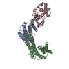 6gn8C 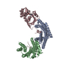 6gnjC  6gnkC 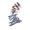 6gnnC 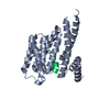 2c23S S: Starting model for refinement C: citing same article ( |
|---|---|
| Similar structure data |


-Links
 PDBj
PDBj


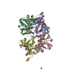
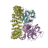
- Assembly
| Deposited unit | 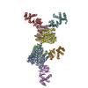
| ||||||||
|---|---|---|---|---|---|---|---|---|---|
| 1 | 
| ||||||||
| 2 | 
| ||||||||
| Unit cell |
|
-Components
| #1: Protein | Mass: 28565.977 Da / Num. of mol.: 4 Source method: isolated from a genetically manipulated source Source: (gene. exp.) Homo sapiens (human) / Gene: YWHAB / Plasmid: pET Duet-1 / Production host: #2: Protein | Mass: 26212.963 Da / Num. of mol.: 4 / Mutation: E379A, E381A Source method: isolated from a genetically manipulated source Source: (gene. exp.) Pseudomonas aeruginosa (bacteria) / Gene: exoS / Plasmid: pET Duet-1 / Production host: |
|---|
-Experimental details
-Experiment
| Experiment | Method: X-RAY DIFFRACTION / Number of used crystals: 1 |
|---|
- Sample preparation
| Crystal | Density Matthews: 2.63 Å3/Da / Density % sol: 53.21 % |
|---|---|
| Crystal grow | Temperature: 277 K / Method: vapor diffusion, sitting drop / pH: 5 / Details: 12% PEG 3350, 4% Tacsimate |
-Data collection
| Diffraction | Mean temperature: 100 K |
|---|---|
| Diffraction source | Source: SYNCHROTRON / Site: ESRF  / Beamline: MASSIF-3 / Wavelength: 0.9677 Å / Beamline: MASSIF-3 / Wavelength: 0.9677 Å |
| Detector | Type: DECTRIS EIGER X 4M / Detector: PIXEL / Date: Dec 11, 2016 |
| Radiation | Protocol: SINGLE WAVELENGTH / Monochromatic (M) / Laue (L): M / Scattering type: x-ray |
| Radiation wavelength | Wavelength: 0.9677 Å / Relative weight: 1 |
| Reflection | Resolution: 3.24→118.1 Å / Num. all: 254184 / Num. obs: 37506 / % possible obs: 99.8 % / Redundancy: 13.3 % / Biso Wilson estimate: 52.97 Å2 / CC1/2: 0.98 / Rrim(I) all: 0.505 / Net I/σ(I): 5 |
| Reflection shell | Resolution: 3.24→3.29 Å / Redundancy: 14 % / Mean I/σ(I) obs: 1.4 / Num. unique obs: 1875 / CC1/2: 0.53 / Rrim(I) all: 2.396 / % possible all: 96.7 |
- Processing
| Software |
| ||||||||||||||||||||||||||||||||||||||||||||||||||||||||||||||||||||||||||||||||||||||||||||||||||||||||||||||||||
|---|---|---|---|---|---|---|---|---|---|---|---|---|---|---|---|---|---|---|---|---|---|---|---|---|---|---|---|---|---|---|---|---|---|---|---|---|---|---|---|---|---|---|---|---|---|---|---|---|---|---|---|---|---|---|---|---|---|---|---|---|---|---|---|---|---|---|---|---|---|---|---|---|---|---|---|---|---|---|---|---|---|---|---|---|---|---|---|---|---|---|---|---|---|---|---|---|---|---|---|---|---|---|---|---|---|---|---|---|---|---|---|---|---|---|---|
| Refinement | Method to determine structure: MOLECULAR REPLACEMENT Starting model: 2C23 Resolution: 3.24→118.05 Å / Cor.coef. Fo:Fc: 0.871 / Cor.coef. Fo:Fc free: 0.813 / Cross valid method: THROUGHOUT / σ(F): 0 / SU Rfree Blow DPI: 0.557
| ||||||||||||||||||||||||||||||||||||||||||||||||||||||||||||||||||||||||||||||||||||||||||||||||||||||||||||||||||
| Displacement parameters | Biso mean: 54.42 Å2
| ||||||||||||||||||||||||||||||||||||||||||||||||||||||||||||||||||||||||||||||||||||||||||||||||||||||||||||||||||
| Refine analyze | Luzzati coordinate error obs: 0.5 Å | ||||||||||||||||||||||||||||||||||||||||||||||||||||||||||||||||||||||||||||||||||||||||||||||||||||||||||||||||||
| Refinement step | Cycle: 1 / Resolution: 3.24→118.05 Å
| ||||||||||||||||||||||||||||||||||||||||||||||||||||||||||||||||||||||||||||||||||||||||||||||||||||||||||||||||||
| Refine LS restraints |
| ||||||||||||||||||||||||||||||||||||||||||||||||||||||||||||||||||||||||||||||||||||||||||||||||||||||||||||||||||
| LS refinement shell | Resolution: 3.24→3.33 Å / Total num. of bins used: 19
|
