 Movie
Movie Controller

[English] 日本語
 Yorodumi
- PDB-3dw8: Structure of a Protein Phosphatase 2A Holoenzyme with B55 subunit -
+ Open data

- Basic information
| Entry | Database: PDB / ID: 3dw8 | |||||||||
|---|---|---|---|---|---|---|---|---|---|---|
| Title | Structure of a Protein Phosphatase 2A Holoenzyme with B55 subunit | |||||||||
 Components Components |
| |||||||||
 Keywords Keywords | HYDROLASE/HYDROLASE INHIBITOR / holoenzyme / B55 / PR55 / WD repeat / Hydrolase / Iron / Manganese / Metal-binding / Methylation / Phosphoprotein / Protein phosphatase / HYDROLASE-HYDROLASE INHIBITOR COMPLEX | |||||||||
| Function / homology |  Function and homology information Function and homology informationregulation of chromosome segregation / meiotic spindle elongation / PP2A-mediated dephosphorylation of key metabolic factors / RNA polymerase II CTD heptapeptide repeat S2 phosphatase activity / RNA polymerase II CTD heptapeptide repeat S7 phosphatase activity / peptidyl-threonine dephosphorylation / regulation of meiotic cell cycle process involved in oocyte maturation / mitotic sister chromatid separation / MASTL Facilitates Mitotic Progression / protein phosphatase type 2A complex ...regulation of chromosome segregation / meiotic spindle elongation / PP2A-mediated dephosphorylation of key metabolic factors / RNA polymerase II CTD heptapeptide repeat S2 phosphatase activity / RNA polymerase II CTD heptapeptide repeat S7 phosphatase activity / peptidyl-threonine dephosphorylation / regulation of meiotic cell cycle process involved in oocyte maturation / mitotic sister chromatid separation / MASTL Facilitates Mitotic Progression / protein phosphatase type 2A complex / meiotic sister chromatid cohesion, centromeric / INTAC complex / RNA polymerase II CTD heptapeptide repeat S5 phosphatase activity / FAR/SIN/STRIPAK complex / female meiotic nuclear division / Regulation of glycolysis by fructose 2,6-bisphosphate metabolism / Inhibition of replication initiation of damaged DNA by RB1/E2F1 / protein phosphatase regulator activity / protein antigen binding / GABA receptor binding / APC truncation mutants have impaired AXIN binding / AXIN missense mutants destabilize the destruction complex / Truncations of AMER1 destabilize the destruction complex / positive regulation of extrinsic apoptotic signaling pathway in absence of ligand / ERKs are inactivated / Initiation of Nuclear Envelope (NE) Reformation / Beta-catenin phosphorylation cascade / Signaling by GSK3beta mutants / CTNNB1 S33 mutants aren't phosphorylated / CTNNB1 S37 mutants aren't phosphorylated / CTNNB1 S45 mutants aren't phosphorylated / CTNNB1 T41 mutants aren't phosphorylated / Co-stimulation by CD28 / regulation of growth / RNA polymerase II transcription initiation surveillance / Disassembly of the destruction complex and recruitment of AXIN to the membrane / protein dephosphorylation / negative regulation of epithelial to mesenchymal transition / Co-inhibition by CTLA4 / Platelet sensitization by LDL / protein-serine/threonine phosphatase / response to morphine / ERK/MAPK targets / negative regulation of glycolytic process through fructose-6-phosphate / vascular endothelial cell response to oscillatory fluid shear stress / protein serine/threonine phosphatase activity / T cell homeostasis / mesoderm development / positive regulation of NLRP3 inflammasome complex assembly / regulation of cell differentiation / regulation of microtubule polymerization / regulation of G1/S transition of mitotic cell cycle / lateral plasma membrane / chromosome, centromeric region / DARPP-32 events / enzyme-substrate adaptor activity / negative regulation of hippo signaling / Cyclin A/B1/B2 associated events during G2/M transition / Nonsense Mediated Decay (NMD) enhanced by the Exon Junction Complex (EJC) / spindle assembly / phosphoprotein phosphatase activity / Amplification of signal from unattached kinetochores via a MAD2 inhibitory signal / Loss of Nlp from mitotic centrosomes / Loss of proteins required for interphase microtubule organization from the centrosome / Recruitment of mitotic centrosome proteins and complexes / protein tyrosine phosphatase activity / Recruitment of NuMA to mitotic centrosomes / Anchoring of the basal body to the plasma membrane / Mitotic Prometaphase / EML4 and NUDC in mitotic spindle formation / protein phosphatase 2A binding / Turbulent (oscillatory, disturbed) flow shear stress activates signaling by PIEZO1 and integrins in endothelial cells / AURKA Activation by TPX2 / Resolution of Sister Chromatid Cohesion / negative regulation of phosphatidylinositol 3-kinase/protein kinase B signal transduction / meiotic cell cycle / chromosome segregation / negative regulation of canonical Wnt signaling pathway / RAF activation / RHO GTPases Activate Formins / Spry regulation of FGF signaling / PKR-mediated signaling / response to lead ion / Degradation of beta-catenin by the destruction complex / tau protein binding / spindle pole / Cyclin D associated events in G1 / Negative regulation of MAPK pathway / Separation of Sister Chromatids / Regulation of TP53 Degradation / Regulation of PLK1 Activity at G2/M Transition / mitotic cell cycle / PI5P, PP2A and IER3 Regulate PI3K/AKT Signaling / microtubule cytoskeleton / protein-containing complex assembly / neuron projection / intracellular signal transduction / membrane raft / protein heterodimerization activity / neuronal cell body Similarity search - Function | |||||||||
| Biological species |  Homo sapiens (human) Homo sapiens (human) Cyanobacteria (cyanobacteria) Cyanobacteria (cyanobacteria) | |||||||||
| Method |  X-RAY DIFFRACTION / SYNCHROTRON / MOLECULAR REPLACEMENT / Resolution: 2.85 Å X-RAY DIFFRACTION / SYNCHROTRON / MOLECULAR REPLACEMENT / Resolution: 2.85 Å | |||||||||
 Authors Authors | Xu, Y. / Chen, Y. / Zhang, P. / Jeffrey, P.D. / Shi, Y. | |||||||||
 Citation Citation | Journal: Mol.Cell / Year: 2008 Title: Structure of a protein phosphatase 2A holoenzyme: insights into B55-mediated Tau dephosphorylation. Authors: Xu, Y. / Chen, Y. / Zhang, P. / Jeffrey, P.D. / Shi, Y. | |||||||||
| History |
|
- Structure visualization
| Structure viewer | Molecule: MolmilJmol/JSmol |
|---|
- Downloads & links
-Download
| PDBx/mmCIF format | 3dw8.cif.gz | 524 KB | Display | PDBx/mmCIF format |
|---|---|---|---|---|
| PDB format | pdb3dw8.ent.gz | 419.7 KB | Display | PDB format |
| PDBx/mmJSON format | 3dw8.json.gz | Tree view | PDBx/mmJSON format | |
| Others |  Other downloads Other downloads |
-Validation report
| Arichive directory | https://data.pdbj.org/pub/pdb/validation_reports/dw/3dw8ftp://data.pdbj.org/pub/pdb/validation_reports/dw/3dw8 | HTTPS FTP |
|---|
-Related structure data
| Similar structure data |
|---|


-Links
 PDBj
PDBj


- Assembly
| Deposited unit | 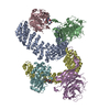
| ||||||||
|---|---|---|---|---|---|---|---|---|---|
| 1 | 
| ||||||||
| 2 | 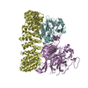
| ||||||||
| Unit cell |
| ||||||||
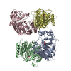
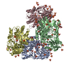
| Details | The biological unit is a heterotrimer. There are two biological units in the asymmetric unit: first heterotrimer consisting of polypeptide chains A,B,C corresponding to PP2A subunits bound to catalytic MN atoms and MCLR inhibitor (chain G), and second heterotrimer consisting of polypeptide chains D,E,F corresponding to PP2A subunits bound to catalytic MN atoms and MCLR inhibitor (chain H). |
-Components
| #1: Protein | Mass: 64762.785 Da / Num. of mol.: 2 / Fragment: A delta 8: Residues 9-589 Source method: isolated from a genetically manipulated source Source: (gene. exp.) Homo sapiens (human) / Gene: PPP2R1A / Production host: #2: Protein | Mass: 51747.984 Da / Num. of mol.: 2 / Mutation: I310V Source method: isolated from a genetically manipulated source Source: (gene. exp.) Homo sapiens (human) / Gene: PPP2R2A / Cell line (production host): SF9 / Production host:   Spodoptera frugiperda (fall armyworm) / References: UniProt: P63151 Spodoptera frugiperda (fall armyworm) / References: UniProt: P63151#3: Protein | Mass: 35636.152 Da / Num. of mol.: 2 Source method: isolated from a genetically manipulated source Source: (gene. exp.) Homo sapiens (human) / Gene: PPP2CA / Cell line (production host): SF9 / Production host: Spodoptera frugiperda (fall armyworm)References: UniProt: P67775, protein-serine/threonine phosphatase #4: Protein/peptide | 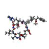  Type: Oligopeptide / Class: Toxin / Mass: 1014.195 Da / Num. of mol.: 2 / Source method: obtained synthetically / Source: (synth.) Cyanobacteria (cyanobacteria) / References: NOR: NOR00109, Microcystin LR Type: Oligopeptide / Class: Toxin / Mass: 1014.195 Da / Num. of mol.: 2 / Source method: obtained synthetically / Source: (synth.) Cyanobacteria (cyanobacteria) / References: NOR: NOR00109, Microcystin LR#5: Chemical | ChemComp-MN /   Mass: 54.938 Da / Num. of mol.: 4 / Source method: obtained synthetically / Formula: Mn Mass: 54.938 Da / Num. of mol.: 4 / Source method: obtained synthetically / Formula: Mn |
|---|
-Experimental details
-Experiment
| Experiment | Method: X-RAY DIFFRACTION / Number of used crystals: 1 |
|---|
- Sample preparation
| Crystal | Density Matthews: 3.11 Å3/Da / Density % sol: 60.47 % |
|---|---|
| Crystal grow | Temperature: 290 K / Method: vapor diffusion, hanging drop / pH: 5.5 Details: 7-10% PEG 35000, 0.10-0.15 M Sodium citrate pH 5.5, VAPOR DIFFUSION, HANGING DROP, temperature 290K |
-Data collection
| Diffraction | Mean temperature: 100 K |
|---|---|
| Diffraction source | Source: SYNCHROTRON / Site: NSLS  / Beamline: X29A / Wavelength: 1.0809 Å / Beamline: X29A / Wavelength: 1.0809 Å |
| Detector | Type: ADSC QUANTUM 315 / Detector: CCD / Date: Apr 1, 2007 |
| Radiation | Monochromator: Si(111) / Protocol: SINGLE WAVELENGTH / Monochromatic (M) / Laue (L): M / Scattering type: x-ray |
| Radiation wavelength | Wavelength: 1.0809 Å / Relative weight: 1 |
| Reflection | Resolution: 2.85→100 Å / Num. all: 87353 / Num. obs: 87353 / % possible obs: 98.9 % / Observed criterion σ(I): -3 / Biso Wilson estimate: 65.9 Å2 |
| Reflection shell | Resolution: 2.85→2.95 Å / % possible all: 99.9 |
- Processing
| Software |
| ||||||||||||||||||||||||||||||||||||||||||||||||||||||||||||
|---|---|---|---|---|---|---|---|---|---|---|---|---|---|---|---|---|---|---|---|---|---|---|---|---|---|---|---|---|---|---|---|---|---|---|---|---|---|---|---|---|---|---|---|---|---|---|---|---|---|---|---|---|---|---|---|---|---|---|---|---|---|
| Refinement | Method to determine structure: MOLECULAR REPLACEMENT Starting model: PP2A A subunit, PP2A C subunit, WD40 ensemble Resolution: 2.85→49.63 Å / Rfactor Rfree error: 0.004 / Data cutoff high absF: 2550770.14 / Data cutoff low absF: 0 / Isotropic thermal model: RESTRAINED / Cross valid method: THROUGHOUT / σ(F): 0 / Stereochemistry target values: Engh & Huber / Details: BULK SOLVENT MODEL USED
| ||||||||||||||||||||||||||||||||||||||||||||||||||||||||||||
| Solvent computation | Solvent model: FLAT MODEL / Bsol: 58.0084 Å2 / ksol: 0.3 e/Å3 | ||||||||||||||||||||||||||||||||||||||||||||||||||||||||||||
| Displacement parameters | Biso mean: 93.1 Å2
| ||||||||||||||||||||||||||||||||||||||||||||||||||||||||||||
| Refine analyze |
| ||||||||||||||||||||||||||||||||||||||||||||||||||||||||||||
| Refinement step | Cycle: LAST / Resolution: 2.85→49.63 Å
| ||||||||||||||||||||||||||||||||||||||||||||||||||||||||||||
| Refine LS restraints |
| ||||||||||||||||||||||||||||||||||||||||||||||||||||||||||||
| LS refinement shell | Resolution: 2.85→3.03 Å / Rfactor Rfree error: 0.017 / Total num. of bins used: 6
| ||||||||||||||||||||||||||||||||||||||||||||||||||||||||||||
| Xplor file |
|
