 Movie
Movie Controller

[English] 日本語
 Yorodumi
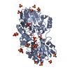
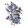
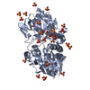
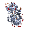
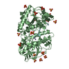
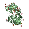
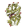
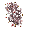
- PDB-3ie0: Crystal Structure of S378Y mutant TTHA0252 from Thermus thermophi... -
+ Open data

- Basic information
| Entry | Database: PDB / ID: 3ie0 | |||||||||
|---|---|---|---|---|---|---|---|---|---|---|
| Title | Crystal Structure of S378Y mutant TTHA0252 from Thermus thermophilus HB8 | |||||||||
 Components Components | Ribonuclease TTHA0252 | |||||||||
 Keywords Keywords | HYDROLASE / Metallo beta lactamase fold / Structural Genomics / NPPSFA / National Project on Protein Structural and Functional Analyses / RIKEN Structural Genomics/Proteomics Initiative / RSGI / Endonuclease / Metal-binding / Nuclease / RNA-binding / rRNA processing | |||||||||
| Function / homology |  Function and homology information Function and homology informationRNA endonuclease activity / rRNA processing / Hydrolases; Acting on ester bonds / hydrolase activity / RNA binding / metal ion binding / cytoplasm Similarity search - Function | |||||||||
| Biological species |   Thermus thermophilus (bacteria) Thermus thermophilus (bacteria) | |||||||||
| Method |  X-RAY DIFFRACTION / SYNCHROTRON / MOLECULAR REPLACEMENT / Resolution: 2.73 Å X-RAY DIFFRACTION / SYNCHROTRON / MOLECULAR REPLACEMENT / Resolution: 2.73 Å | |||||||||
 Authors Authors | Ishikawa, H. / Nakagawa, N. / Kuramitsu, S. / Yokoyama, S. / Masui, R. / RIKEN Structural Genomics/Proteomics Initiative (RSGI) | |||||||||
 Citation Citation | Journal: To be Published Title: Crystal Structure of S378Y mutant TTHA0252 from Thermus thermophilus HB8 Authors: Ishikawa, H. / Nakagawa, N. / Kuramitsu, S. / Masui, R. | |||||||||
| History |
|
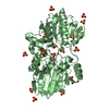
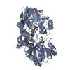
- Structure visualization
| Structure viewer | Molecule: MolmilJmol/JSmol |
|---|
- Downloads & links
-Download
| PDBx/mmCIF format | 3ie0.cif.gz | 342.7 KB | Display | PDBx/mmCIF format |
|---|---|---|---|---|
| PDB format | pdb3ie0.ent.gz | 281.5 KB | Display | PDB format |
| PDBx/mmJSON format | 3ie0.json.gz | Tree view | PDBx/mmJSON format | |
| Others |  Other downloads Other downloads |
-Validation report
| Arichive directory | https://data.pdbj.org/pub/pdb/validation_reports/ie/3ie0ftp://data.pdbj.org/pub/pdb/validation_reports/ie/3ie0 | HTTPS FTP |
|---|
-Related structure data
| Related structure data |  2dkfS S: Starting model for refinement |
|---|---|
| Similar structure data | |
| Other databases |


-Links
 PDBj
PDBj


- Assembly
| Deposited unit | 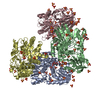
| ||||||||
|---|---|---|---|---|---|---|---|---|---|
| 1 | 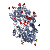
| ||||||||
| 2 | 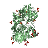
| ||||||||
| 3 | 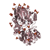
| ||||||||
| 4 | 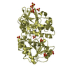
| ||||||||
| Unit cell |
|
-Components
| #1: Protein | Mass: 47194.168 Da / Num. of mol.: 4 / Mutation: S378Y Source method: isolated from a genetically manipulated source Source: (gene. exp.) Thermus thermophilus (bacteria) / Strain: HB8 / Gene: TTHA0252 / Plasmid: pET11a / Production host: References: UniProt: Q5SLP1, Hydrolases; Acting on ester bonds #2: Chemical | ChemComp-SO4 /   Mass: 96.063 Da / Num. of mol.: 67 / Source method: obtained synthetically / Formula: SO4 Mass: 96.063 Da / Num. of mol.: 67 / Source method: obtained synthetically / Formula: SO4#3: Chemical |   Mass: 189.100 Da / Num. of mol.: 3 / Source method: obtained synthetically / Formula: C6H5O7 Mass: 189.100 Da / Num. of mol.: 3 / Source method: obtained synthetically / Formula: C6H5O7#4: Chemical | ChemComp-ZN /   Mass: 65.409 Da / Num. of mol.: 8 / Source method: obtained synthetically / Formula: Zn Mass: 65.409 Da / Num. of mol.: 8 / Source method: obtained synthetically / Formula: Zn#5: Water | ChemComp-HOH / |  Mass: 18.015 Da / Num. of mol.: 80 / Source method: isolated from a natural source / Formula: H2O Mass: 18.015 Da / Num. of mol.: 80 / Source method: isolated from a natural source / Formula: H2O |
|---|
-Experimental details
-Experiment
| Experiment | Method: X-RAY DIFFRACTION / Number of used crystals: 1 |
|---|
- Sample preparation
| Crystal | Density Matthews: 3.16 Å3/Da / Density % sol: 61.08 % / Mosaicity: 0.266 ° |
|---|---|
| Crystal grow | Temperature: 293 K / Method: vapor diffusion, hanging drop / pH: 5.6 Details: 50mM sodium citrate, 0.2M ammonium sulfate, 0.6M lithium sulfate, pH 5.6, VAPOR DIFFUSION, HANGING DROP, temperature 293K |
-Data collection
| Diffraction | Mean temperature: 95 K | |||||||||||||||||||||||||||||||||||||||||||||||||||||||||||||||||||||||||||||
|---|---|---|---|---|---|---|---|---|---|---|---|---|---|---|---|---|---|---|---|---|---|---|---|---|---|---|---|---|---|---|---|---|---|---|---|---|---|---|---|---|---|---|---|---|---|---|---|---|---|---|---|---|---|---|---|---|---|---|---|---|---|---|---|---|---|---|---|---|---|---|---|---|---|---|---|---|---|---|
| Diffraction source | Source: SYNCHROTRON / Site: SPring-8  / Beamline: BL44B2 / Wavelength: 1.282245 Å / Beamline: BL44B2 / Wavelength: 1.282245 Å | |||||||||||||||||||||||||||||||||||||||||||||||||||||||||||||||||||||||||||||
| Detector | Type: ADSC QUANTUM 210 / Detector: CCD / Date: Jul 14, 2007 | |||||||||||||||||||||||||||||||||||||||||||||||||||||||||||||||||||||||||||||
| Radiation | Monochromator: transparent diamond double crystal / Protocol: SINGLE WAVELENGTH / Monochromatic (M) / Laue (L): M / Scattering type: x-ray | |||||||||||||||||||||||||||||||||||||||||||||||||||||||||||||||||||||||||||||
| Radiation wavelength | Wavelength: 1.282245 Å / Relative weight: 1 | |||||||||||||||||||||||||||||||||||||||||||||||||||||||||||||||||||||||||||||
| Reflection | Resolution: 2.73→50 Å / Num. obs: 61873 / % possible obs: 99.5 % / Observed criterion σ(I): -3 / Redundancy: 3.8 % / Rmerge(I) obs: 0.044 / Χ2: 1.065 / Net I/σ(I): 15.1 | |||||||||||||||||||||||||||||||||||||||||||||||||||||||||||||||||||||||||||||
| Reflection shell |
|
- Processing
| Software |
| ||||||||||||||||||||||||||||||||||||
|---|---|---|---|---|---|---|---|---|---|---|---|---|---|---|---|---|---|---|---|---|---|---|---|---|---|---|---|---|---|---|---|---|---|---|---|---|---|
| Refinement | Method to determine structure: MOLECULAR REPLACEMENT Starting model: 2DKF Resolution: 2.73→50 Å / Occupancy max: 1 / Occupancy min: 1 / Isotropic thermal model: restrained / Cross valid method: THROUGHOUT / σ(F): 0 / Stereochemistry target values: Engh & Huber
| ||||||||||||||||||||||||||||||||||||
| Solvent computation | Bsol: 38.53 Å2 | ||||||||||||||||||||||||||||||||||||
| Displacement parameters | Biso max: 178.75 Å2 / Biso mean: 64.278 Å2 / Biso min: 12.36 Å2
| ||||||||||||||||||||||||||||||||||||
| Refine analyze |
| ||||||||||||||||||||||||||||||||||||
| Refinement step | Cycle: LAST / Resolution: 2.73→50 Å
| ||||||||||||||||||||||||||||||||||||
| Refine LS restraints |
| ||||||||||||||||||||||||||||||||||||
| LS refinement shell | Resolution: 2.73→2.9 Å / Rfactor Rfree error: 0.013
| ||||||||||||||||||||||||||||||||||||
| Xplor file |
|
