 Movie
Movie Controller

[English] 日本語
 Yorodumi
- PDB-6gci: Structure of the bongkrekic acid-inhibited mitochondrial ADP/ATP ... -
+ Open data

- Basic information
| Entry | Database: PDB / ID: 6gci | ||||||||||||
|---|---|---|---|---|---|---|---|---|---|---|---|---|---|
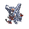
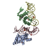
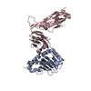
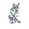
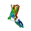
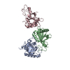
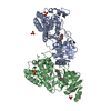
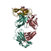
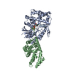
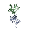
| Title | Structure of the bongkrekic acid-inhibited mitochondrial ADP/ATP carrier | ||||||||||||
 Components Components |
| ||||||||||||
 Keywords Keywords | MEMBRANE PROTEIN / Transporter / Inhibitor / Mitochondrial / Carrier | ||||||||||||
| Function / homology |  Function and homology information Function and homology informationmitochondrial ADP transmembrane transport / ATP:ADP antiporter activity / mitochondrial ATP transmembrane transport / mitochondrial inner membrane Similarity search - Function | ||||||||||||
| Biological species | Thermothelomyces thermophila | ||||||||||||
| Method |  X-RAY DIFFRACTION / SYNCHROTRON / MOLECULAR REPLACEMENT / Resolution: 3.3 Å X-RAY DIFFRACTION / SYNCHROTRON / MOLECULAR REPLACEMENT / Resolution: 3.3 Å | ||||||||||||
 Authors Authors | Ruprecht, J.J. / King, M.S. / Pardon, E. / Aleksandrova, A.A. / Zogg, T. / Crichton, P.G. / Steyaert, J. / Kunji, E.R.S. | ||||||||||||
| Funding support |  United Kingdom, United Kingdom,  Belgium, 3items Belgium, 3items
| ||||||||||||
 Citation Citation | Journal: Cell / Year: 2019 Title: The Molecular Mechanism of Transport by the Mitochondrial ADP/ATP Carrier. Authors: Ruprecht, J.J. / King, M.S. / Zogg, T. / Aleksandrova, A.A. / Pardon, E. / Crichton, P.G. / Steyaert, J. / Kunji, E.R.S. | ||||||||||||
| History |
|
- Structure visualization
| Structure viewer | Molecule: MolmilJmol/JSmol |
|---|
- Downloads & links
-Download
| PDBx/mmCIF format | 6gci.cif.gz | 189.7 KB | Display | PDBx/mmCIF format |
|---|---|---|---|---|
| PDB format | pdb6gci.ent.gz | 147.2 KB | Display | PDB format |
| PDBx/mmJSON format | 6gci.json.gz | Tree view | PDBx/mmJSON format | |
| Others |  Other downloads Other downloads |
-Validation report
| Arichive directory | https://data.pdbj.org/pub/pdb/validation_reports/gc/6gciftp://data.pdbj.org/pub/pdb/validation_reports/gc/6gci | HTTPS FTP |
|---|
-Related structure data


-Links
 PDBj
PDBj


- Assembly
| Deposited unit | 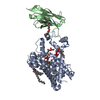
| ||||||||
|---|---|---|---|---|---|---|---|---|---|
| 1 |
| ||||||||
| Unit cell |
| ||||||||
| Components on special symmetry positions |
|
-Components
| #1: Protein | Mass: 34042.578 Da / Num. of mol.: 1 / Mutation: Q302K Source method: isolated from a genetically manipulated source Details: Sequence has Q302K mutation Source: (gene. exp.)  Thermothelomyces thermophila (strain ATCC 42464 / BCRC 31852 / DSM 1799) (fungus) Thermothelomyces thermophila (strain ATCC 42464 / BCRC 31852 / DSM 1799) (fungus)Strain: ATCC 42464 / BCRC 31852 / DSM 1799 / Gene: MYCTH_2316753 / Plasmid: pYES3 / Details (production host): modified / Production host:  | ||||
|---|---|---|---|---|---|
| #2: Antibody | Mass: 14521.907 Da / Num. of mol.: 1 Source method: isolated from a genetically manipulated source Details: Nanobody selected against the BKA-inhibited ADP/ATP carrier Source: (gene. exp.)  | ||||
| #3: Chemical | ChemComp-BKC /   Mass: 486.597 Da / Num. of mol.: 1 / Source method: obtained synthetically / Formula: C28H38O7 Mass: 486.597 Da / Num. of mol.: 1 / Source method: obtained synthetically / Formula: C28H38O7 | ||||
| #4: Chemical | ChemComp-CDL /   Mass: 1464.043 Da / Num. of mol.: 5 / Source method: obtained synthetically / Formula: C81H156O17P2 / Comment: phospholipid*YM Mass: 1464.043 Da / Num. of mol.: 5 / Source method: obtained synthetically / Formula: C81H156O17P2 / Comment: phospholipid*YM#5: Chemical | ChemComp-P6G / |   Mass: 282.331 Da / Num. of mol.: 1 / Source method: obtained synthetically / Formula: C12H26O7 / Comment: precipitant*YM Mass: 282.331 Da / Num. of mol.: 1 / Source method: obtained synthetically / Formula: C12H26O7 / Comment: precipitant*YMHas protein modification | Y | |
-Experimental details
-Experiment
| Experiment | Method: X-RAY DIFFRACTION / Number of used crystals: 1 |
|---|
- Sample preparation
| Crystal | Density Matthews: 5.03 Å3/Da / Density % sol: 75.53 % |
|---|---|
| Crystal grow | Temperature: 283 K / Method: vapor diffusion, sitting drop Details: 0.1M MES pH 6.5, 1% 3-methyl-3-pentanol, 22% PEG400 |
-Data collection
| Diffraction | Mean temperature: 100 K |
|---|---|
| Diffraction source | Source: SYNCHROTRON / Site: Diamond / Beamline: I24 / Wavelength: 0.9686 Å |
| Detector | Type: DECTRIS PILATUS3 6M / Detector: PIXEL / Date: Aug 4, 2017 |
| Radiation | Protocol: SINGLE WAVELENGTH / Monochromatic (M) / Laue (L): M / Scattering type: x-ray |
| Radiation wavelength | Wavelength: 0.9686 Å / Relative weight: 1 |
| Reflection | Resolution: 3.3→29.35 Å / Num. obs: 13204 / % possible obs: 85.1 % / Redundancy: 4.7 % / Biso Wilson estimate: 111.32 Å2 / Rmerge(I) obs: 0.063 / Net I/σ(I): 12.1 |
| Reflection shell | Resolution: 3.3→3.43 Å / Redundancy: 4.7 % / Rmerge(I) obs: 0.688 / Mean I/σ(I) obs: 2.3 / Num. unique obs: 659 / % possible all: 40.3 |
- Processing
| Software |
| ||||||||||||||||||||||||||||||||||||||||||||||||||||||||||||||||||||||||||||||||||||||||||||||||||||||||||||||||||
|---|---|---|---|---|---|---|---|---|---|---|---|---|---|---|---|---|---|---|---|---|---|---|---|---|---|---|---|---|---|---|---|---|---|---|---|---|---|---|---|---|---|---|---|---|---|---|---|---|---|---|---|---|---|---|---|---|---|---|---|---|---|---|---|---|---|---|---|---|---|---|---|---|---|---|---|---|---|---|---|---|---|---|---|---|---|---|---|---|---|---|---|---|---|---|---|---|---|---|---|---|---|---|---|---|---|---|---|---|---|---|---|---|---|---|---|
| Refinement | Method to determine structure: MOLECULAR REPLACEMENT Starting model: 5IMK, 4C9H Resolution: 3.3→29.35 Å / Cor.coef. Fo:Fc: 0.869 / Cor.coef. Fo:Fc free: 0.889 / Cross valid method: THROUGHOUT / σ(F): 0 / SU Rfree Blow DPI: 0.484 / SU Rfree Cruickshank DPI: 0.487 Details: (A) Crystal diffracted anisotropically, and data was treated with STARANISO. (B) Fourier map coefficients have been deposited along with the observed data. (C) A PEG molecule lies on a ...Details: (A) Crystal diffracted anisotropically, and data was treated with STARANISO. (B) Fourier map coefficients have been deposited along with the observed data. (C) A PEG molecule lies on a symmetry axis (special position) and has been modelled with occupancy=0.5. (D) Some cardiolipins molecules have been modelled with partial occupancy, as described in the accompanying paper. (E) Met250-Ser252 and Lys257 lie in very weak electron density. Their position is supported by good electron density obtained from an earlier crystal. (F) There are unmodelled bits of electron density near the N-terminus of the carrier, which are likely to be lipid headgroups/tails. (G) Additional electron density at the C-terminus of the carrier indicates where the C-terminal tail is likely to run, but it is not possible to model this accurately. (H) The structure factor file contains a second data block containing the structure factors from a crystal of BKA-inhibited TtAac grown without the nanobody. This P212121 crystal diffracted to lower resolution and with significant anisotropy, but was used to confirm the domain positions observed in the P3221 crystal of the TtAac-Nb complex. Full details can be found in the primary citation.
| ||||||||||||||||||||||||||||||||||||||||||||||||||||||||||||||||||||||||||||||||||||||||||||||||||||||||||||||||||
| Displacement parameters | Biso mean: 111.34 Å2
| ||||||||||||||||||||||||||||||||||||||||||||||||||||||||||||||||||||||||||||||||||||||||||||||||||||||||||||||||||
| Refine analyze | Luzzati coordinate error obs: 0.55 Å | ||||||||||||||||||||||||||||||||||||||||||||||||||||||||||||||||||||||||||||||||||||||||||||||||||||||||||||||||||
| Refinement step | Cycle: 1 / Resolution: 3.3→29.35 Å
| ||||||||||||||||||||||||||||||||||||||||||||||||||||||||||||||||||||||||||||||||||||||||||||||||||||||||||||||||||
| Refine LS restraints |
| ||||||||||||||||||||||||||||||||||||||||||||||||||||||||||||||||||||||||||||||||||||||||||||||||||||||||||||||||||
| LS refinement shell | Resolution: 3.3→3.57 Å / Total num. of bins used: 7
| ||||||||||||||||||||||||||||||||||||||||||||||||||||||||||||||||||||||||||||||||||||||||||||||||||||||||||||||||||
| Refinement TLS params. | Method: refined / Refine-ID: X-RAY DIFFRACTION
| ||||||||||||||||||||||||||||||||||||||||||||||||||||||||||||||||||||||||||||||||||||||||||||||||||||||||||||||||||
| Refinement TLS group |
|
