 Movie
Movie Controller

[English] 日本語
 Yorodumi
- PDB-3cx7: Crystal Structure of PDZRhoGEF rgRGS Domain in a Complex with Gal... -
+ Open data

- Basic information
| Entry | Database: PDB / ID: 3cx7 | ||||||
|---|---|---|---|---|---|---|---|
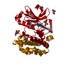
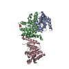
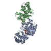
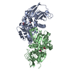
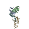
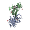
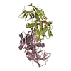
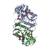
| Title | Crystal Structure of PDZRhoGEF rgRGS Domain in a Complex with Galpha-13 Bound to GDP-AlF4 | ||||||
 Components Components |
| ||||||
 Keywords Keywords | SIGNALING PROTEIN / SIGNAL TRANSDUCTION / PROTEIN COMPLEX | ||||||
| Function / homology |  Function and homology information Function and homology informationSema4D induced cell migration and growth-cone collapse / RHOB GTPase cycle / CDC42 GTPase cycle / NRAGE signals death through JNK / D5 dopamine receptor binding / G alpha (12/13) signalling events / CDC42 GTPase cycle / RAC1 GTPase cycle / NRAGE signals death through JNK / regulation of fibroblast migration ...Sema4D induced cell migration and growth-cone collapse / RHOB GTPase cycle / CDC42 GTPase cycle / NRAGE signals death through JNK / D5 dopamine receptor binding / G alpha (12/13) signalling events / CDC42 GTPase cycle / RAC1 GTPase cycle / NRAGE signals death through JNK / regulation of fibroblast migration / Thromboxane signalling through TP receptor / Rho-activating G protein-coupled receptor signaling pathway / RHOA GTPase cycle / positive regulation of L-glutamate import across plasma membrane / Thrombin signalling through proteinase activated receptors (PARs) / RAC1 GTPase cycle / G alpha (12/13) signalling events / branching involved in blood vessel morphogenesis / negative regulation of vascular associated smooth muscle cell migration / negative regulation of vascular associated smooth muscle cell proliferation / regulation of postsynapse assembly / Rho protein signal transduction / guanyl-nucleotide exchange factor activity / regulation of cell migration / GTPase activator activity / brush border membrane / platelet activation / G protein-coupled receptor binding / regulation of blood pressure / G-protein beta/gamma-subunit complex binding / adenylate cyclase-modulating G protein-coupled receptor signaling pathway / melanosome / regulation of cell shape / heterotrimeric G-protein complex / adenylate cyclase-activating G protein-coupled receptor signaling pathway / positive regulation of cytosolic calcium ion concentration / G protein activity / angiogenesis / in utero embryonic development / cell differentiation / intracellular signal transduction / postsynapse / G protein-coupled receptor signaling pathway / GTPase activity / positive regulation of DNA-templated transcription / GTP binding / nucleoplasm / metal ion binding / nucleus / plasma membrane / cytoplasm / cytosol Similarity search - Function | ||||||
| Biological species |  | ||||||
| Method |  X-RAY DIFFRACTION / SYNCHROTRON / Resolution: 2.25 Å X-RAY DIFFRACTION / SYNCHROTRON / Resolution: 2.25 Å | ||||||
 Authors Authors | Sprang, S.R. / Chen, Z. | ||||||
 Citation Citation | Journal: Structure / Year: 2008 Title: Recognition of the Activated States of Galpha13 by the rgRGS Domain of PDZRhoGEF. Authors: Chen, Z. / Singer, W.D. / Danesh, S.M. / Sternweis, P.C. / Sprang, S.R. | ||||||
| History |
|
- Structure visualization
| Structure viewer | Molecule: MolmilJmol/JSmol |
|---|
- Downloads & links
-Download
| PDBx/mmCIF format | 3cx7.cif.gz | 122.3 KB | Display | PDBx/mmCIF format |
|---|---|---|---|---|
| PDB format | pdb3cx7.ent.gz | 92 KB | Display | PDB format |
| PDBx/mmJSON format | 3cx7.json.gz | Tree view | PDBx/mmJSON format | |
| Others |  Other downloads Other downloads |
-Validation report
| Arichive directory | https://data.pdbj.org/pub/pdb/validation_reports/cx/3cx7ftp://data.pdbj.org/pub/pdb/validation_reports/cx/3cx7 | HTTPS FTP |
|---|
-Related structure data
| Related structure data |  3cx6C 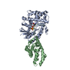 3cx8C 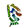 1htjS 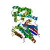 1zcbS C: citing same article ( S: Starting model for refinement |
|---|---|
| Similar structure data |


-Links
 PDBj
PDBj


- Assembly
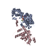
| Deposited unit | 
| ||||||||
|---|---|---|---|---|---|---|---|---|---|
| 1 |
| ||||||||
| Unit cell |
|
-Components
-Protein , 2 types, 2 molecules AB
| #1: Protein | Mass: 39743.516 Da / Num. of mol.: 1 / Fragment: N-terminally truncated Source method: isolated from a genetically manipulated source Source: (gene. exp.)   Spodoptera frugiperda (fall armyworm) / References: UniProt: P27601 Spodoptera frugiperda (fall armyworm) / References: UniProt: P27601 |
|---|---|
| #2: Protein | Mass: 23323.350 Da / Num. of mol.: 1 / Fragment: RhoGEF-RGS (rgRGS) Domain Source method: isolated from a genetically manipulated source Source: (gene. exp.)  |
-Non-polymers , 4 types, 117 molecules 


| #3: Chemical | ChemComp-MG /  Mass: 24.305 Da / Num. of mol.: 1 / Source method: obtained synthetically / Formula: Mg Mass: 24.305 Da / Num. of mol.: 1 / Source method: obtained synthetically / Formula: Mg |
|---|---|
| #4: Chemical | ChemComp-ALF /  Mass: 102.975 Da / Num. of mol.: 1 / Source method: obtained synthetically / Formula: AlF4 Mass: 102.975 Da / Num. of mol.: 1 / Source method: obtained synthetically / Formula: AlF4 |
| #5: Chemical | ChemComp-GDP /  Type: RNA linking / Mass: 443.201 Da / Num. of mol.: 1 / Source method: obtained synthetically / Formula: C10H15N5O11P2 / Comment: GDP, energy-carrying molecule*YM Type: RNA linking / Mass: 443.201 Da / Num. of mol.: 1 / Source method: obtained synthetically / Formula: C10H15N5O11P2 / Comment: GDP, energy-carrying molecule*YM |
| #6: Water | ChemComp-HOH / Mass: 18.015 Da / Num. of mol.: 114 / Source method: isolated from a natural source / Formula: H2O |
-Experimental details
-Experiment
| Experiment | Method: X-RAY DIFFRACTION / Number of used crystals: 1 |
|---|
- Sample preparation
| Crystal | Density Matthews: 2.31 Å3/Da / Density % sol: 46.64 % |
|---|---|
| Crystal grow | Temperature: 293 K / Method: vapor diffusion, hanging drop / pH: 7.1 Details: 18-23 % polyethylene glycol monomethyl ether 2000 and 100 mM Tris, pH 6.9-7.3, pH 7.1, VAPOR DIFFUSION, HANGING DROP, temperature 293K |
-Data collection
| Diffraction | Mean temperature: 100 K |
|---|---|
| Diffraction source | Source: SYNCHROTRON / Site: APS  / Beamline: 23-ID-D / Wavelength: 0.97939 Å / Beamline: 23-ID-D / Wavelength: 0.97939 Å |
| Detector | Type: ADSC QUANTUM 315 / Detector: CCD / Date: Jun 24, 2007 / Details: Mirrors |
| Radiation | Monochromator: SAGITALLY FOCUSED Si(111) / Protocol: SINGLE WAVELENGTH / Monochromatic (M) / Laue (L): M / Scattering type: x-ray |
| Radiation wavelength | Wavelength: 0.97939 Å / Relative weight: 1 |
| Reflection | Resolution: 2.25→45 Å / Num. obs: 26672 / % possible obs: 93.6 % / Observed criterion σ(F): 1 / Observed criterion σ(I): 1 / Redundancy: 5.9 % / Biso Wilson estimate: 37.2 Å2 / Rsym value: 0.086 / Net I/σ(I): 25.6 |
| Reflection shell | Resolution: 2.25→2.3 Å / Redundancy: 2.2 % / Mean I/σ(I) obs: 2 / Rsym value: 0.411 / % possible all: 60.2 |
- Processing
| Software |
| ||||||||||||||||||||||||||||
|---|---|---|---|---|---|---|---|---|---|---|---|---|---|---|---|---|---|---|---|---|---|---|---|---|---|---|---|---|---|
| Refinement | Starting model: PDB Entry 1ZCB, 1HTJ Resolution: 2.25→45 Å / Isotropic thermal model: Isotropic / Cross valid method: THROUGHOUT / σ(F): 0 / σ(I): 0 / Stereochemistry target values: Engh & Huber
| ||||||||||||||||||||||||||||
| Solvent computation | Bsol: 35.979 Å2 | ||||||||||||||||||||||||||||
| Displacement parameters | Biso mean: 42.307 Å2
| ||||||||||||||||||||||||||||
| Refinement step | Cycle: LAST / Resolution: 2.25→45 Å
| ||||||||||||||||||||||||||||
| Refine LS restraints |
| ||||||||||||||||||||||||||||
| Xplor file |
|
