[English] 日本語
Yorodumi
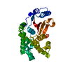
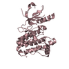
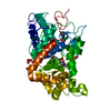
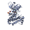
- PDB-3pyy: Discovery and Characterization of a Cell-Permeable, Small-molecul... -

+
Open data


ID or keywords:

Loading...

-
Basic information

Entry
Database: PDB / ID: 3pyy
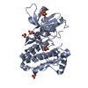
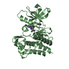
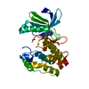
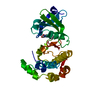
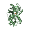
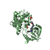
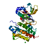
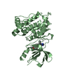
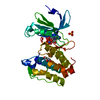
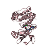
TitleDiscovery and Characterization of a Cell-Permeable, Small-molecule c-Abl Kinase Activator that Binds to the Myristoyl Binding Site
ComponentsTyrosine-protein kinase ABL1
KeywordsTRANSFERASE / Tyrosine kinase
Function / homology
Function and homology information


protein localization to cytoplasmic microtubule plus-end / DNA conformation change / mitochondrial depolarization / response to epinephrine / phospholipase C-inhibiting G protein-coupled receptor signaling pathway / negative regulation of ubiquitin-protein transferase activity / podocyte apoptotic process / regulation of postsynaptic specialization assembly / positive regulation of phospholipase C/protein kinase C signal transduction / regulation of modification of synaptic structure ...protein localization to cytoplasmic microtubule plus-end / DNA conformation change / mitochondrial depolarization / response to epinephrine / phospholipase C-inhibiting G protein-coupled receptor signaling pathway / negative regulation of ubiquitin-protein transferase activity / podocyte apoptotic process / regulation of postsynaptic specialization assembly / positive regulation of phospholipase C/protein kinase C signal transduction / regulation of modification of synaptic structure / nicotinate-nucleotide adenylyltransferase activity / delta-catenin binding / Role of ABL in ROBO-SLIT signaling / positive regulation of extracellular matrix organization / neuropilin signaling pathway / neuropilin binding / regulation of cell motility / bubble DNA binding / positive regulation of establishment of T cell polarity / regulation of T cell differentiation / positive regulation of blood vessel branching / proline-rich region binding / cellular response to dopamine / positive regulation of dendrite development / mitogen-activated protein kinase binding / regulation of Cdc42 protein signal transduction / regulation of hematopoietic stem cell differentiation / syntaxin binding / regulation of axon extension / positive regulation of cell migration involved in sprouting angiogenesis / Myogenesis / HDR through Single Strand Annealing (SSA) / platelet-derived growth factor receptor-beta signaling pathway / RUNX2 regulates osteoblast differentiation / Fc-gamma receptor signaling pathway involved in phagocytosis / vascular endothelial cell response to oscillatory fluid shear stress / myoblast proliferation / regulation of endocytosis / cardiac muscle cell proliferation / regulation of microtubule polymerization / negative regulation of long-term synaptic potentiation / associative learning / positive regulation of focal adhesion assembly / actin monomer binding / cellular response to transforming growth factor beta stimulus / ephrin receptor signaling pathway / positive regulation of vasoconstriction / regulation of cell adhesion / positive regulation of substrate adhesion-dependent cell spreading / endothelial cell migration / positive regulation of stress fiber assembly / RHO GTPases Activate WASPs and WAVEs / negative regulation of double-strand break repair via homologous recombination / positive regulation of T cell migration / mismatch repair / ephrin receptor binding / four-way junction DNA binding / ruffle / signal transduction in response to DNA damage / phosphotyrosine residue binding / actin filament polymerization / positive regulation of endothelial cell migration / SH2 domain binding / integrin-mediated signaling pathway / protein serine/threonine kinase activator activity / positive regulation of fibroblast proliferation / response to endoplasmic reticulum stress / Turbulent (oscillatory, disturbed) flow shear stress activates signaling by PIEZO1 and integrins in endothelial cells / protein kinase C binding / protein modification process / regulation of actin cytoskeleton organization / intrinsic apoptotic signaling pathway in response to DNA damage / non-specific protein-tyrosine kinase / FCGR3A-mediated phagocytosis / non-membrane spanning protein tyrosine kinase activity / Regulation of actin dynamics for phagocytic cup formation / regulation of autophagy / cellular response to hydrogen peroxide / epidermal growth factor receptor signaling pathway / enzyme activator activity / autophagy / positive regulation of neuron apoptotic process / sequence-specific double-stranded DNA binding / kinase activity / Cyclin D associated events in G1 / actin filament binding / actin cytoskeleton organization / actin cytoskeleton / manganese ion binding / mitotic cell cycle / positive regulation of cytosolic calcium ion concentration / nuclear membrane / Recruitment and ATM-mediated phosphorylation of repair and signaling proteins at DNA double strand breaks / Factors involved in megakaryocyte development and platelet production / MLL4 and MLL3 complexes regulate expression of PPARG target genes in adipogenesis and hepatic steatosis / growth cone / RUNX1 regulates transcription of genes involved in differentiation of HSCs / protein tyrosine kinase activity / response to oxidative stress / cellular response to oxidative stress
Similarity search - Function
F-actin binding / F-actin binding / F-actin binding domain (FABD) / Tyrosine-protein kinase ABL, SH2 domain / : / SH3 domain / SH2 domain / Src homology 2 (SH2) domain profile. / Src homology 2 domains / SH2 domain ...F-actin binding / F-actin binding / F-actin binding domain (FABD) / Tyrosine-protein kinase ABL, SH2 domain / : / SH3 domain / SH2 domain / Src homology 2 (SH2) domain profile. / Src homology 2 domains / SH2 domain / Src homology 3 domains / SH2 domain superfamily / SH3-like domain superfamily / Src homology 3 (SH3) domain profile. / SH3 domain / Tyrosine-protein kinase, catalytic domain / Tyrosine kinase, catalytic domain / Tyrosine protein kinases specific active-site signature. / Tyrosine-protein kinase, active site / Protein tyrosine and serine/threonine kinase / Serine-threonine/tyrosine-protein kinase, catalytic domain / Phosphorylase Kinase; domain 1 / Phosphorylase Kinase; domain 1 / Transferase(Phosphotransferase) domain 1 / Transferase(Phosphotransferase); domain 1 / Protein kinase, ATP binding site / Protein kinases ATP-binding region signature. / Protein kinase domain profile. / Protein kinase domain / Protein kinase-like domain superfamily / 2-Layer Sandwich / Orthogonal Bundle / Mainly Alpha / Alpha Beta
Similarity search - Domain/homology
Chem-3YY / Chem-STI / Tyrosine-protein kinase ABL1
Similarity search - Component
Biological speciesHomo sapiens (human)
MethodX-RAY DIFFRACTION / SYNCHROTRON / MOLECULAR REPLACEMENT / Resolution: 1.85 Å
AuthorsYang, J. / Campobasso, N. / Biju, M.P. / Fisher, K. / Pan, X.Q. / Cottom, J. / Galbraith, S. / Ho, T. / Zhang, H. / Hong, X. ...Yang, J. / Campobasso, N. / Biju, M.P. / Fisher, K. / Pan, X.Q. / Cottom, J. / Galbraith, S. / Ho, T. / Zhang, H. / Hong, X. / Ward, P. / Hofmann, G. / Siegfried, B.
CitationJournal: Chem.Biol. / Year: 2011
Title: Discovery and Characterization of a Cell-Permeable, Small-Molecule c-Abl Kinase Activator that Binds to the Myristoyl Binding Site.
Authors: Yang, J. / Campobasso, N. / Biju, M.P. / Fisher, K. / Pan, X.Q. / Cottom, J. / Galbraith, S. / Ho, T. / Zhang, H. / Hong, X. / Ward, P. / Hofmann, G. / Siegfried, B. / Zappacosta, F. / ...Authors: Yang, J. / Campobasso, N. / Biju, M.P. / Fisher, K. / Pan, X.Q. / Cottom, J. / Galbraith, S. / Ho, T. / Zhang, H. / Hong, X. / Ward, P. / Hofmann, G. / Siegfried, B. / Zappacosta, F. / Washio, Y. / Cao, P. / Qu, J. / Bertrand, S. / Wang, D.Y. / Head, M.S. / Li, H. / Moores, S. / Lai, Z. / Johanson, K. / Burton, G. / Erickson-Miller, C. / Simpson, G. / Tummino, P. / Copeland, R.A. / Oliff, A.
History
DepositionDec 13, 2010Deposition site: RCSB / Processing site: RCSB
Revision 1.0Mar 9, 2011Provider: repository / Type: Initial release
Revision 1.1Jul 13, 2011Group: Version format compliance
Revision 1.2Jan 24, 2018Group: Structure summary / Category: audit_author / Item: _audit_author.name
Revision 1.3Feb 21, 2024Group: Data collection / Database references ...Data collection / Database references / Derived calculations / Structure summary
Category: chem_comp / chem_comp_atom ...chem_comp / chem_comp_atom / chem_comp_bond / database_2 / struct_ref_seq_dif / struct_site
Item: _chem_comp.pdbx_synonyms / _database_2.pdbx_DOI ..._chem_comp.pdbx_synonyms / _database_2.pdbx_DOI / _database_2.pdbx_database_accession / _struct_ref_seq_dif.details / _struct_site.pdbx_auth_asym_id / _struct_site.pdbx_auth_comp_id / _struct_site.pdbx_auth_seq_id
Revision 1.4May 7, 2025Group: Database references / Source and taxonomy / Structure summary
Category: entity / entity_name_com ...entity / entity_name_com / entity_src_gen / pdbx_entry_details / struct_ref / struct_ref_seq / struct_ref_seq_dif
Item: _entity.pdbx_description / _entity.pdbx_ec ..._entity.pdbx_description / _entity.pdbx_ec / _entity_src_gen.pdbx_beg_seq_num / _entity_src_gen.pdbx_end_seq_num / _entity_src_gen.pdbx_gene_src_gene / _entity_src_gen.pdbx_seq_type / _struct_ref.db_code / _struct_ref.pdbx_align_begin / _struct_ref.pdbx_db_accession / _struct_ref_seq.db_align_beg / _struct_ref_seq.db_align_end / _struct_ref_seq.pdbx_db_accession / _struct_ref_seq_dif.pdbx_seq_db_accession_code

-
Structure visualization

Structure viewerMolecule:
MolmilJmol/JSmol

Downloads & links

-
Assembly

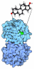
Deposited unit
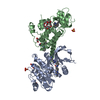
A: Tyrosine-protein kinase ABL1
B: Tyrosine-protein kinase ABL1
hetero molecules


Theoretical massNumber of molelcules
Total (without water)71,87211
Polymers69,4172
Non-polymers2,4559
Water3,873215
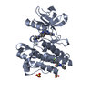
1
A: Tyrosine-protein kinase ABL1
hetero molecules


Theoretical massNumber of molelcules
Total (without water)35,7315
Polymers34,7091
Non-polymers1,0224
Water181
TypeNameSymmetry operationNumber
identity operation1_555x,y,z1
MethodPISA
2
B: Tyrosine-protein kinase ABL1
hetero molecules


Theoretical massNumber of molelcules
Total (without water)36,1416
Polymers34,7091
Non-polymers1,4335
Water181
TypeNameSymmetry operationNumber
identity operation1_555x,y,z1
MethodPISA
Unit cell
Length a, b, c (Å)74.391, 95.432, 115.502
Angle α, β, γ (deg.)90.00, 90.00, 90.00
Int Tables number19
Space group name H-MP212121

-
Components

-
Protein , 1 types, 2 molecules AB

#1: Protein Tyrosine-protein kinase ABL1 / Abelson murine leukemia viral oncogene homolog 1 / Abelson tyrosine-protein kinase 1 / Proto- ...Abelson murine leukemia viral oncogene homolog 1 / Abelson tyrosine-protein kinase 1 / Proto-oncogene c-Abl / p150


Mass: 34708.566 Da / Num. of mol.: 2 / Fragment: UNP residues 266-549
Source method: isolated from a genetically manipulated source
Source: (gene. exp.) Homo sapiens (human) / Gene: ABL1, ABL, JTK7 / Production host: Spodoptera frugiperda (fall armyworm)
References: UniProt: P00519, non-specific protein-tyrosine kinase

-
Non-polymers , 6 types, 224 molecules

#2: Chemical ChemComp-SO4 / SULFATE ION


Mass: 96.063 Da / Num. of mol.: 3 / Source method: obtained synthetically / Formula: SO4
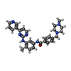
#3: Chemical ChemComp-STI / 4-(4-METHYL-PIPERAZIN-1-YLMETHYL)-N-[4-METHYL-3-(4-PYRIDIN-3-YL-PYRIMIDIN-2-YLAMINO)-PHENYL]-BENZAMIDE / STI-571 / IMATINIB


Mass: 493.603 Da / Num. of mol.: 2 / Source method: obtained synthetically / Formula: C29H31N7O / Comment: medication*YM
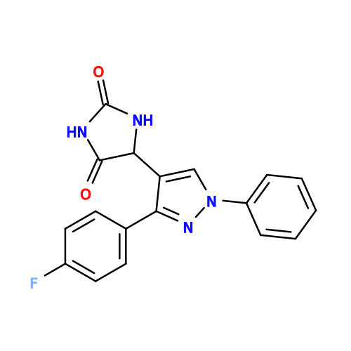
#4: Chemical ChemComp-3YY / (5R)-5-[3-(4-fluorophenyl)-1-phenyl-1H-pyrazol-4-yl]imidazolidine-2,4-dione


Mass: 336.320 Da / Num. of mol.: 2 / Source method: obtained synthetically / Formula: C18H13FN4O2
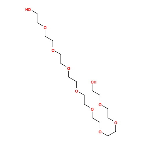
#5: Chemical ChemComp-2PE / NONAETHYLENE GLYCOL


Mass: 414.488 Da / Num. of mol.: 1 / Source method: obtained synthetically / Formula: C18H38O10 / Comment: precipitant*YM
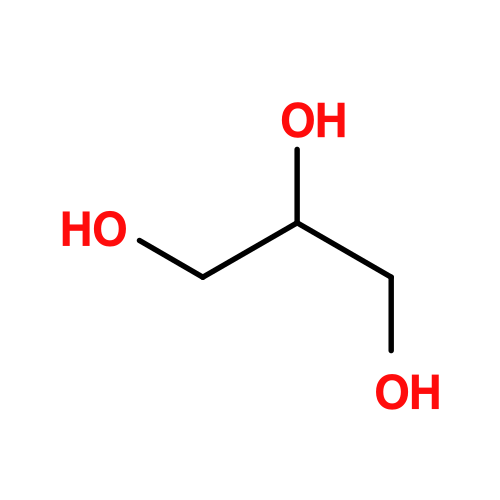
#6: Chemical ChemComp-GOL / GLYCEROL / GLYCERIN / PROPANE-1,2,3-TRIOL


Mass: 92.094 Da / Num. of mol.: 1 / Source method: obtained synthetically / Formula: C3H8O3
#7: Water ChemComp-HOH / water


Mass: 18.015 Da / Num. of mol.: 215 / Source method: isolated from a natural source / Formula: H2O

-
Details

Has protein modificationN

-
Experimental details

-
Experiment

ExperimentMethod: X-RAY DIFFRACTION / Number of used crystals: 1

-
Sample preparation

CrystalDensity Matthews: 2.95 Å3/Da / Density % sol: 58.35 %
Crystal growTemperature: 277 K / Method: vapor diffusion, hanging drop / pH: 8
Details: 20 mgs/ml protein in 20 mM Tris-HCl, pH 8.0, 100 mM NaCl, 3 mM DTT and 5% (v/v) glycerol. Reservoir with 0.4 M ammonium phosphate. Cryo w/ 15%-30% glycerol, VAPOR DIFFUSION, HANGING DROP, temperature 277K

-
Data collection

DiffractionMean temperature: 100 K
Diffraction sourceSource: SYNCHROTRON / Site: APS / Beamline: 21-ID-G / Wavelength: 0.97872 Å
DetectorType: RAYONIX MX-225 / Detector: CCD / Date: Feb 17, 2010
RadiationMonochromator: diamond / Protocol: SINGLE WAVELENGTH / Monochromatic (M) / Laue (L): M / Scattering type: x-ray
Radiation wavelengthWavelength: 0.97872 Å / Relative weight: 1
ReflectionResolution: 1.85→30 Å / Num. obs: 69872 / % possible obs: 99 % / Observed criterion σ(F): 1 / Observed criterion σ(I): 1

-
Processing

Software
NameVersionClassification
MAR345dtbdata collection
PHASERphasing
PHENIX(phenix.refine: 1.6_289)refinement
HKL-2000data reduction
SCALEPACKdata scaling
RefinementMethod to determine structure: MOLECULAR REPLACEMENT / Resolution: 1.85→29.963 Å / SU ML: 0.18 / σ(F): 0.03 / Stereochemistry target values: ML
RfactorNum. reflection% reflectionSelection details
Rfree0.2039 1922 2.87 %Random
Rwork0.1886 ---
obs0.1891 66950 94.89 %-
Solvent computationShrinkage radii: 0.9 Å / VDW probe radii: 1.11 Å / Solvent model: FLAT BULK SOLVENT MODEL / Bsol: 60.008 Å2 / ksol: 0.4 e/Å3
Displacement parameters
Baniso -1Baniso -2Baniso -3
1-2.378 Å20 Å2-0 Å2
2--3.617 Å2-0 Å2
3----5.995 Å2
Refinement stepCycle: LAST / Resolution: 1.85→29.963 Å
ProteinNucleic acidLigandSolventTotal
Num. atoms4223 0 173 215 4611
Refine LS restraints
Refine-IDTypeDev idealNumber
X-RAY DIFFRACTIONf_bond_d0.0074528
X-RAY DIFFRACTIONf_angle_d1.0766125
X-RAY DIFFRACTIONf_dihedral_angle_d15.8361620
X-RAY DIFFRACTIONf_chiral_restr0.071631
X-RAY DIFFRACTIONf_plane_restr0.004803
LS refinement shell
Resolution (Å)Rfactor RfreeNum. reflection RfreeRfactor RworkNum. reflection RworkRefine-ID% reflection obs (%)
1.85-1.89860.24451190.23493972X-RAY DIFFRACTION82
1.8986-1.94990.2461220.22154149X-RAY DIFFRACTION86
1.9499-2.00730.24411310.2044400X-RAY DIFFRACTION91
2.0073-2.0720.221320.19764482X-RAY DIFFRACTION93
2.072-2.14610.20531380.1894598X-RAY DIFFRACTION95
2.1461-2.2320.22061340.18174604X-RAY DIFFRACTION95
2.232-2.33350.221370.18824692X-RAY DIFFRACTION96
2.3335-2.45650.21391400.18714728X-RAY DIFFRACTION97
2.4565-2.61030.221400.1914772X-RAY DIFFRACTION98
2.6103-2.81170.19081420.194808X-RAY DIFFRACTION98
2.8117-3.09440.23691460.20364873X-RAY DIFFRACTION99
3.0944-3.54160.17971440.19274900X-RAY DIFFRACTION99
3.5416-4.45960.18481460.16554940X-RAY DIFFRACTION99
4.4596-29.96720.19321510.18485110X-RAY DIFFRACTION99

+
About Yorodumi

-
News

-
Feb 9, 2022. New format data for meta-information of EMDB entries

New format data for meta-information of EMDB entries

  • Version 3 of the EMDB header file is now the official format.
  • The previous official version 1.9 will be removed from the archive.

Related info.:EMDB header

External links:wwPDB to switch to version 3 of the EMDB data model

-
Aug 12, 2020. Covid-19 info

Covid-19 info

URL: https://pdbj.org/emnavi/covid19.php

New page: Covid-19 featured information page in EM Navigator.

Related info.:Covid-19 info / Mar 5, 2020. Novel coronavirus structure data

+
Mar 5, 2020. Novel coronavirus structure data

Novel coronavirus structure data

Related info.:Yorodumi Speices / Aug 12, 2020. Covid-19 info

External links:COVID-19 featured content - PDBj / Molecule of the Month (242):Coronavirus Proteases

+
Jan 31, 2019. EMDB accession codes are about to change! (news from PDBe EMDB page)

EMDB accession codes are about to change! (news from PDBe EMDB page)

  • The allocation of 4 digits for EMDB accession codes will soon come to an end. Whilst these codes will remain in use, new EMDB accession codes will include an additional digit and will expand incrementally as the available range of codes is exhausted. The current 4-digit format prefixed with “EMD-” (i.e. EMD-XXXX) will advance to a 5-digit format (i.e. EMD-XXXXX), and so on. It is currently estimated that the 4-digit codes will be depleted around Spring 2019, at which point the 5-digit format will come into force.
  • The EM Navigator/Yorodumi systems omit the EMD- prefix.

Related info.:Q: What is EMD? / ID/Accession-code notation in Yorodumi/EM Navigator

External links:EMDB Accession Codes are Changing Soon! / Contact to PDBj

+
Jul 12, 2017. Major update of PDB

Major update of PDB

  • wwPDB released updated PDB data conforming to the new PDBx/mmCIF dictionary.
  • This is a major update changing the version number from 4 to 5, and with Remediation, in which all the entries are updated.
  • In this update, many items about electron microscopy experimental information are reorganized (e.g. em_software).
  • Now, EM Navigator and Yorodumi are based on the updated data.

External links:wwPDB Remediation / Enriched Model Files Conforming to OneDep Data Standards Now Available in the PDB FTP Archive

-
Yorodumi

Thousand views of thousand structures

  • Yorodumi is a browser for structure data from EMDB, PDB, SASBDB, etc.
  • This page is also the successor to EM Navigator detail page, and also detail information page/front-end page for Omokage search.
  • The word "yorodu" (or yorozu) is an old Japanese word meaning "ten thousand". "mi" (miru) is to see.

Related info.:EMDB / PDB / SASBDB / Comparison of 3 databanks / Yorodumi Search / Aug 31, 2016. New EM Navigator & Yorodumi / Yorodumi Papers / Jmol/JSmol / Function and homology information / Changes in new EM Navigator and Yorodumi

Read more